Inserting part of endoscope

A technology of endoscope and insertion part, which is applied in the field of medical devices, can solve the problems of increased chance of eyeball infection, decreased eyeball tolerance, and damage to eyeball wall integrity, so as to reduce the chance of infection, improve the success rate and reduce the risk Effect

Inactive Publication Date: 2010-03-17
卿国平
View PDF0 Cites 2 Cited by
  • Summary
  • Abstract
  • Description
  • Claims
  • Application Information

AI Technical Summary

Problems solved by technology

This operation achieves the purpose of drainage by cutting through the wall of the eyeball. Cutting through the wall of the eyeball not only destroys the integrity of the eyeball wall, but also greatly increases the chance of eyeball infection, and the ability of the eyeball to withstand external trauma after surgery is reduced

Method used


Image

  • Inserting part of endoscope
  • Inserting part of endoscope
  • Inserting part of endoscope

Examples


Embodiment 1

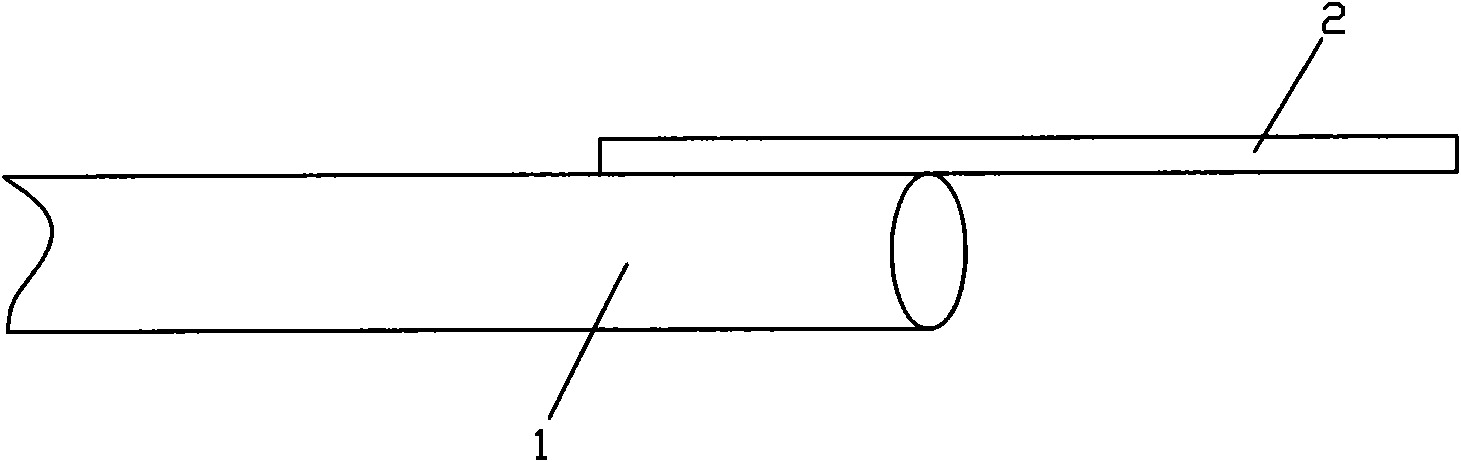
[0024] Such as Figure 1a and 1b As shown, the endoscope insertion part of the present invention includes an endoscope 1 and a surgical tool, wherein the surgical tool is a straight strip-shaped rigid long rod 2, one end of which is connected to the surface or front of the endoscope 1, and the other end faces toward the endoscope 1. Extending forward, the endoscope 1 in the present invention is a conventional soft endoscope or hard endoscope, and its internal lighting device provides a light source that can illuminate the inspection and operation range including surgical tools; the other end is the operation end, Direct implementation of separation, displacement or foreign body removal functions during surgical operations.

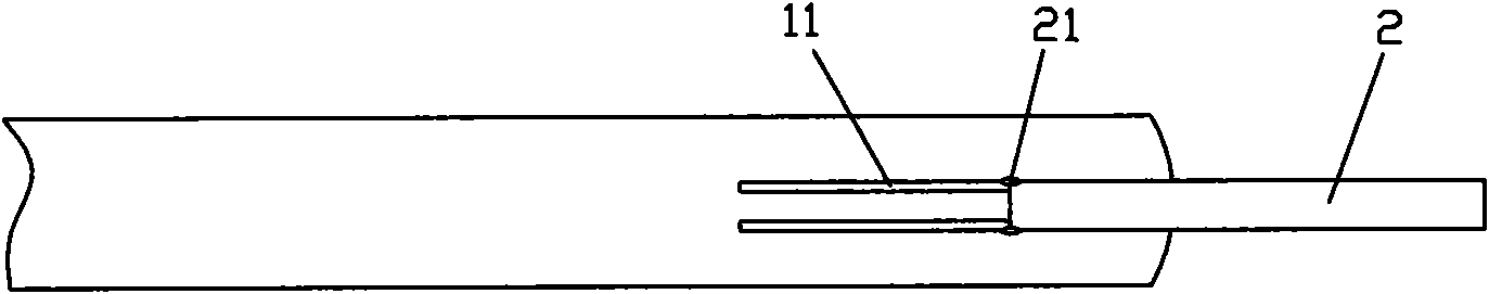
[0025] The connecting end of the surgical tool and the endoscope 1 can be hinged or fixedly connected. In order to facilitate the surgical incision where the endoscope 1 passes through the cornea, the endoscope 1 is provided with a slideway 11, and the sur...

Embodiment 2

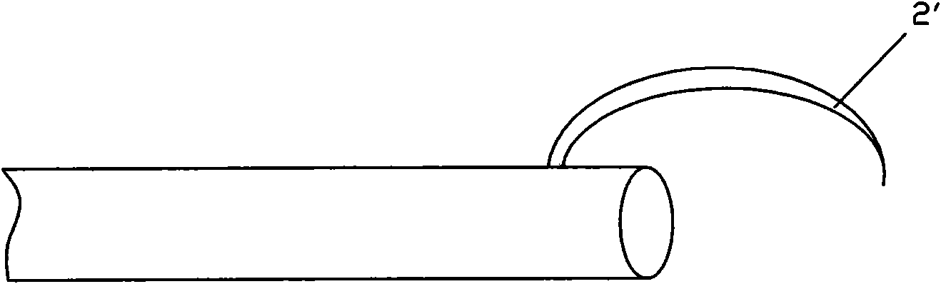
[0027] Such as Figure 1c As shown, the shape of the surgical tool of the endoscope insertion part of the present invention is an arc rod 2 ', and other parts are the same as the endoscope insertion part in Embodiment 1.

Embodiment 3

[0029] Such as Figure 1d As shown, the shape of the surgical tool of the endoscope insertion part of the present invention is a bending rod 2 ", and other parts are the same as the endoscope insertion part in Embodiment 1.


PUM


Abstract

The invention discloses an inserting part of an endoscope, comprising an endoscope body and an operation tool installed on the endoscope body. The operation tool is hard, and has the functions of bluntly separating, moving and actually cutting on a tissue structure in a projection scope of an optical lighting system in the endoscope body. The inserting part of the endoscope can be extended into the angle of the anterior chamber of an eye through a cornea cut to conduct operations such as separating, cutting and foreign matter-taking out on the angle of the anterior chamber and the recess of achamber angle, thereby separating the chamber angle under observation by the endoscope, greatly improving the availability and safety of closed-angle glaucoma, and providing a new choice for treatingforeign matter in chamber angle.

Description

technical field [0001] The invention relates to the field of medical instruments, in particular to an endoscope insertion part. Background technique [0002] Glaucoma is a group of eye diseases characterized by the death of retinal ganglion cells caused by an absolute or relative increase in intraocular pressure, and the common clinical manifestations are increased intraocular pressure, visual field defect, pallor of the retina, and enlarged cup-to-disk ratio. Glaucoma is a common and frequently-occurring disease in ophthalmology. Because of the irreversible characteristics of blindness, most of the patients are chronic and the symptoms are hidden. Most patients are not discovered until the late stage or after blindness in one eye, which brings difficulties to the diagnosis and treatment of glaucoma. Glaucoma is currently one of the leading causes of blindness worldwide. [0003] Glaucoma is divided into open-angle type and angle-closure type according to the state of the c...

Claims


Application Information

Patent Timeline
Patent Type & Authority Applications(China)
IPC IPC(8): A61B3/117A61F9/007
Inventor 卿国平
Owner 卿国平
Why Eureka
Try Eureka