
Method for recognizing function response signal under function nuclear magnetic resonance scan

A functional nuclear magnetic resonance and response signal technology, which is applied in the direction of using nuclear magnetic resonance spectrum for measurement, magnetic resonance measurement, and measurement using nuclear magnetic resonance imaging system, etc. problem, to achieve the effect of eliminating the influence of noise

Inactive Publication Date: 2013-06-05
SOUTHEAST UNIV +1
View PDF0 Cites 0 Cited by
  • Summary
  • Abstract
  • Description
  • Claims
  • Application Information

AI Technical Summary

Problems solved by technology

The currently widely used method is to use the hemodynamic response function (HRF) of the blood oxygen saturation concentration (BOLD) signal. This method relies on the existing fixed model and assumptions on the signal distribution, and it is difficult to find differences between individuals. Discrimination against non-functional signals is also not robust enough
In recent years, data-driven methods have also been developed to obtain functional response signals (the independent component fractal method proposed by Martin is the most typical, see Martin J.M., Scott M., Greg G.B.et.al, Analysis of fMRI Data by Blind Separation into Independent Spatial Components, Human Brain Mapping, 1998, 6: 160-188), but the method itself can only decompose each disordered signal component, and the brain function signal can only be distinguished by the shape of the component wave
In view of this, these methods are more suitable for data with typical functional response signal waveforms, such as the experimental data under the block design, but they are powerless for the experimental data under the event-related design, which is more and more widely used recently

Method used


Image

  • Method for recognizing function response signal under function nuclear magnetic resonance scan
  • Method for recognizing function response signal under function nuclear magnetic resonance scan
  • Method for recognizing function response signal under function nuclear magnetic resonance scan

Examples


Embodiment Construction

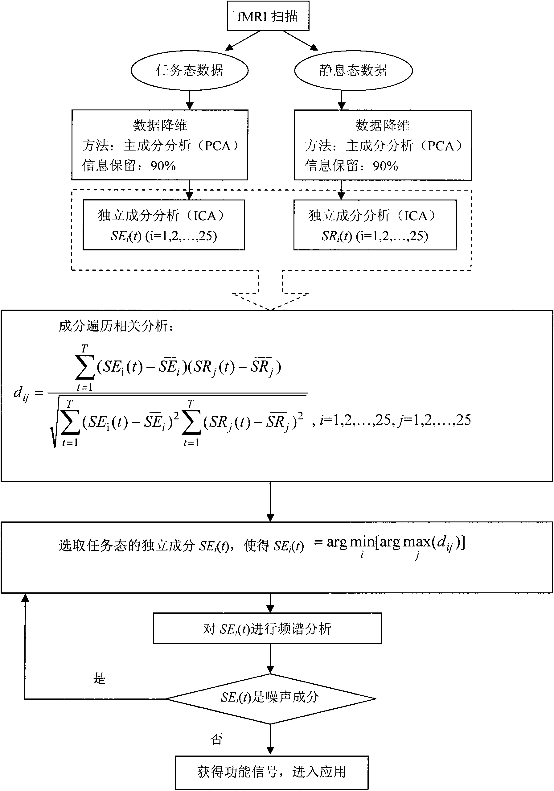
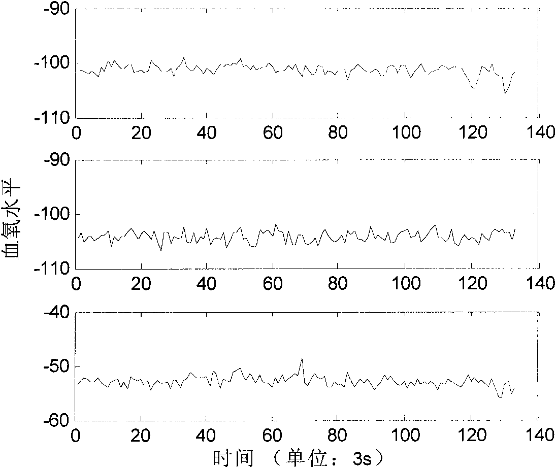
[0028] Under the stimulation of the Chinese Facial Expression Video System (CFEVS), the subjects scanned to obtain task state data, and then scanned to obtain rest state data. The fMRI analysis software SPM was used to preprocess the data spatially and temporally, including head movement correction, spatial normalization, Gaussian smoothing filtering, time normalization and high-pass filtering to remove low-frequency noise, and after operation, the data set was obtained. Use PCA (Tipping M, BishopC.Mixtures of probabilistic principal component analyzers.NeuralComputation, 1999, 11: 443-482) to reduce the dimension of task state data and rest state data, and retain the main information; then use the time domain Independent component analysis (ICA) extracts 25 independent components of each of the two types of data; and then finds out the range of functional signal components by performing correlation traversal between the independent components of the two types of data, and obta...


PUM


Abstract

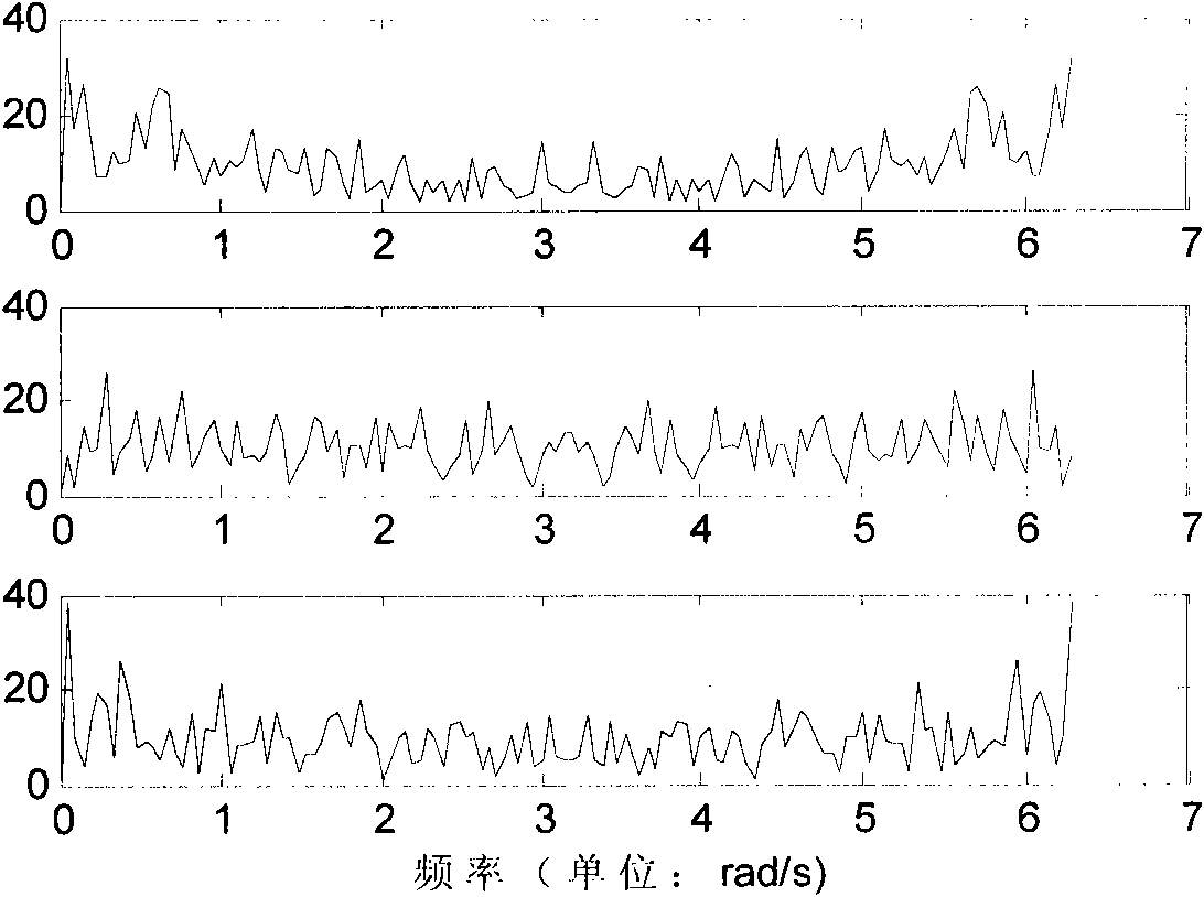
The invention relates to a method for recognizing a function response signal under function nuclear magnetic resonance scan, comprising the following steps of: (1) obtaining task-state data and resting-state data by utilizing a nuclear magnetic resonance apparatus, processing the data in space and time by utilizing a function nuclear magnetic resonance analysis software SPM (statistical parametric mapping) to obtain a data set; (2)reducing the dimensionality of the task-state data and the resting-state data by utilizing a current principal component analysis PCA method, conserving main information, i.e. conserving the eigenvectors of information energy of over 90 percent of the data, reestablishing data, and then respectively extracting independent components of the two kinds of data including a machine noise signal component, a non-neurogenic physiological noise component and a neurogenic function signal response component by utilizing an independent component analysis ICA method in current time domain; (3) finding out the range of the function signal component by carrying out corresponding traversal among independent components of the two kinds of data to obtain a data set containing the function signal component; and (4) carrying out spectral analysis on every signal in the data set, eliminating components without obvious energy peak values in frequency domain, and selecting a principal component from rest components which is the principal function respond signal.

Description

technical field [0001] The invention relates to the extraction of functional nuclear magnetic resonance (fMRI) signals, in particular to a screening method for functional response signals under functional nuclear magnetic resonance (fMRI) scanning, and belongs to the technical fields of image processing and frequency spectrum analysis. Background technique [0002] Functional magnetic resonance (fMRI) signals are very sensitive. During the acquisition of time images, physiological signals caused by the subject's tiny head movements, heart beats, eye movements and some irrelevant muscle movements are also mixed in the fMRI signals. Constitutes the noise signal in the brain function signal. In order to extract the functional response signals under external stimuli, these noise signals must be separated from the brain function signals. The currently widely used method is to use the hemodynamic response function (HRF) of the blood oxygen saturation concentration (BOLD) signal. ...

Claims


Application Information

Patent Timeline
Patent Type & Authority Patents(China)
IPC IPC(8): A61B5/055G01R33/46G01R33/465G01R33/56
Inventor 卢青姚志剑刘刚
Owner SOUTHEAST UNIV
Learn More