Canny model-based method for segmenting three-dimensional medical image
A medical image and image technology, applied in the field of image processing
Inactive Publication Date: 2010-09-08
UNIV OF ELECTRONICS SCI & TECH OF CHINA
View PDF2 Cites 20 Cited by
- Summary
- Abstract
- Description
- Claims
- Application Information
AI Technical Summary
Problems solved by technology
[0005] However, due to the differences in the imaging principles of medical images and the characteristics of the tissues themselves, and the formation of images is subject to factors such as noise, field offset effects, local body effects, and tissue motion, between tissues, between tissues and organs, and between organs and organs. It is very necessary to quickly and accurately realize 3D medical image segmentation
Method used
View moreImage
Smart ImageViewing Examples
Examples
Embodiment Construction
[0047] When the technical solution of the present invention is realized, first use the Matlab language to write the program; then use the three-dimensional MRI or CT medical image data to perform parameter setting and program optimization processing; finally use the C++ language to rewrite the program code and the interactive interface framework to improve program performance .
Login to View More PUM
 Abstract
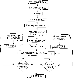
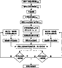
The invention relates to a Canny model-based method for segmenting a three-dimensional medical image and belongs to the technical field of image processing. The method comprises the following steps of: capturing a three-dimensional section image Imnk comprising a user-interested target in an original three-dimensional medical image IMNK through user interaction; performing median filtering on the three-dimensional section image Imnk to remove image noise; acquiring a Canny edge information image Cmnk of the three-dimensional section image Imnk which is subjected to the median filtering by a Canny method; searching a target edge and extracting a complete enclosed target edge according to interaction point coordinates (i0, j0) near the target edge of a certain frame of image of the user; and extracting interested target edges Emnk of all frames in the three-dimensional section image Imnk, wherein the extracted interested target edges Emnk serve as a segmentation result of the three-dimensional medical image. The Canny model-based method for segmenting the three-dimensional medical image uses a few user interaction processes, has a small calculated amount and can rapidly and effectively extract edge information of interested targets in the three-dimensional medical image so as to complete the segmentation of the three-dimensional medical image.
Description
technical field [0001] The invention belongs to the technical field of image processing, and relates to an interactive fast segmentation method of three-dimensional medical images. Background technique [0002] In order to accurately distinguish normal tissue structures and abnormal lesions in medical images, medical images need to be segmented. Traditional image segmentation methods mainly include: [0003] (1) Edge-based segmentation methods: usually use different properties between regions (such as gray discontinuity in the region) to divide the boundaries between regions, such methods include parallel differential operator methods (such as Roberts, Sobel, Laplacian, Marr and other operators), serial boundary search methods, methods based on surface fitting, etc.; [0004] (2) Region-based segmentation methods: Usually, the homogeneity within the same region is used to identify different regions in an image, including threshold methods, region growing and splitting and ...
Claims
Login to View More Application Information
Patent Timeline
 Patent Type & Authority Applications(China)
IPC IPC(8): G06T7/00
Inventor 解梅吴炳荣
Owner UNIV OF ELECTRONICS SCI & TECH OF CHINA
Features
- R&D
- Intellectual Property
- Life Sciences
- Materials
- Tech Scout
- Unparalleled Data Quality
- Higher Quality Content
- 60% Fewer Hallucinations
Learn More Browse by: Latest US Patents, China's latest patents, Technical Efficacy Thesaurus, Application Domain, Technology Topic, Popular Technical Reports.
© 2025 PatSnap. All rights reserved.Legal|Privacy policy|Modern Slavery Act Transparency Statement|Sitemap|About US| Contact US: help@patsnap.com


