
Rapid X-ray fluorescence CT method

An X-ray and fluorescence technology, applied in the field of X-ray imaging, can solve the problems of restricting wide application and taking a long time, and achieve the effects of simplifying experimental equipment, improving data acquisition speed, and easy correction

Inactive Publication Date: 2010-10-20
SHANGHAI INST OF APPLIED PHYSICS - CHINESE ACAD OF SCI
View PDF0 Cites 31 Cited by
  • Summary
  • Abstract
  • Description
  • Claims
  • Application Information

AI Technical Summary

Problems solved by technology

Due to the point scanning method used in data collection, it takes a long time to obtain a set of two-dimensional or three-dimensional experimental data, which limits the wide application of this method

Method used


Image

  • Rapid X-ray fluorescence CT method
  • Rapid X-ray fluorescence CT method
  • Rapid X-ray fluorescence CT method

Examples


Embodiment Construction

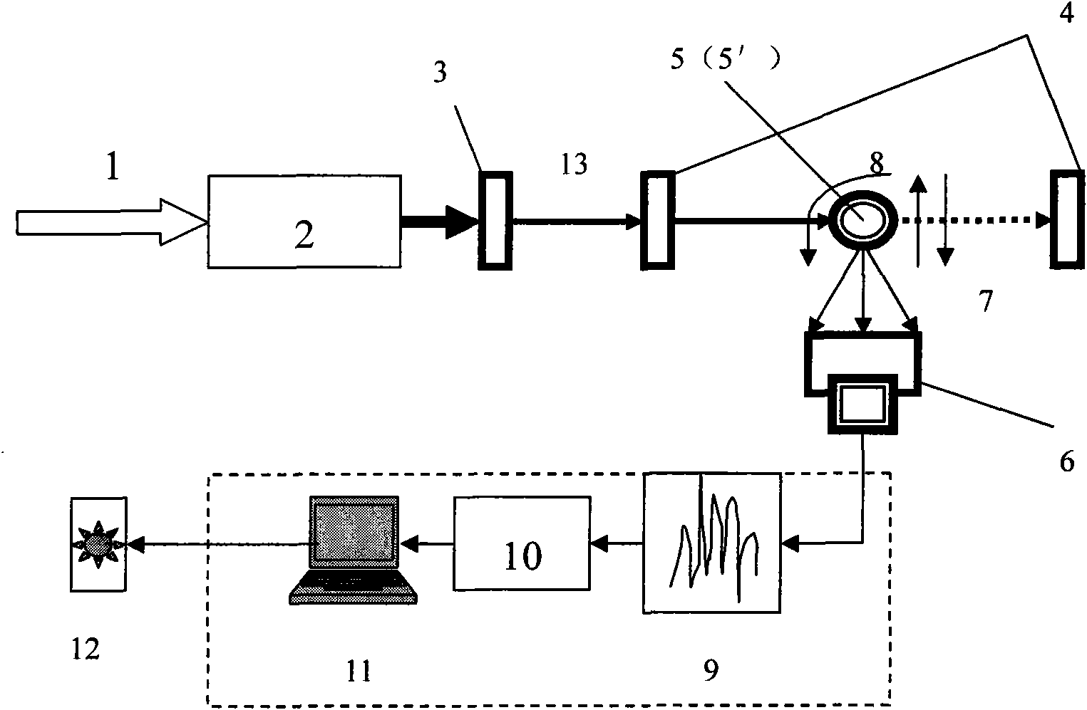
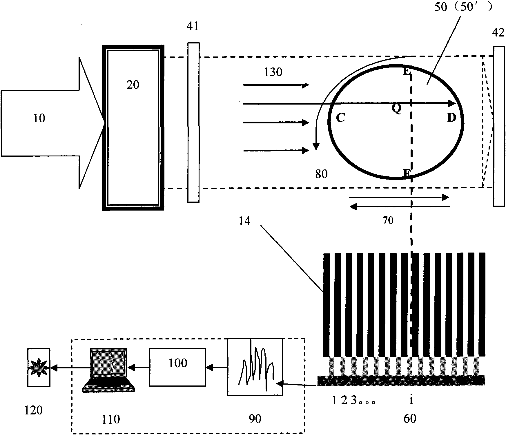
[0018] Attached below image 3 Specific embodiments of the present invention are given.

[0019] In the fast X-ray fluorescence CT experiment according to the present invention, adopt such as image 3 A diagram of the experimental setup is shown. The experimental equipment mainly includes a monochromator 20 , two X-ray intensity detectors 41 and 42 , an array fluorescence detector 60 equipped with a lead collimator 14 , a sample stage 50 and data processing systems 90 , 100 , and 110 .

[0020] The operating principle of the fast X-ray fluorescence CT method according to the present invention is as follows:

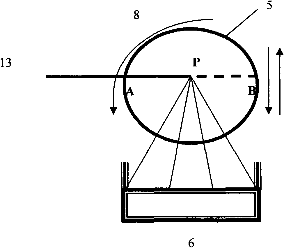
[0021] The synchrotron radiation light 10 is monochromated by the monochromator 20, and the formed monochromatic large spot 130 is directly irradiated on the sample 50' without passing through the beam limitation of the micro-beam device, and the characteristic X-ray fluorescence inside the sample 50' is excited, and the above-mentioned fluorescence is Recorded by the ...


PUM


Abstract

The invention improves the data acquisition speed of traditional X-ray fluorescence CT. The technical scheme is to provide a rapid X-ray fluorescence CT method which comprises the following steps: using a large light spot to irradiate a sample and using an array fluorescence detector with a lead collimator to record fluorescence energy spectrum. Compared with the traditional X-ray fluorescence CT method, the data acquisition speed of the method is improved by times. The improved times of the data acquisition speed is equivalent to the increased times of the array cells of the array fluorescence detector. For example, for a 64x64 array fluorescence detector, the data acquisition speed can be improved by 64x64 times. The method can realize rapid and in-situ X-ray fluorescence CT experiments.

Description

technical field [0001] The invention relates to an X-ray imaging method, in particular to a fast X-ray fluorescence CT method. Background technique [0002] X-ray fluorescence computed tomography (CT) method is the product of the organic combination of X-ray CT and X-ray fluorescence analysis. It is a non-destructive testing technology that reconstructs non-radioactive elements (such as calcium , iron, etc.) in the three-dimensional distribution image of the sample, which can simultaneously measure the distribution of multiple elements in the sample without the need for destructive slice processing of the sample. The main experimental equipment such as figure 1 As shown, it mainly includes a monochromator 2, a micro-beam device 3, two X-ray intensity detectors 4, a fluorescence detector 6, a sample stage 5 (rotatable and translational), and data processing systems 9, 10, 11. [0003] In the traditional X-ray fluorescence CT method, the monochromatic X-ray micro-beam 13 is ...

Claims


Application Information

Patent Timeline
Patent Type & Authority Applications(China)
IPC IPC(8): A61B6/03A61B6/00
Inventor 邓彪杨群谢红兰杜国浩肖体乔
Owner SHANGHAI INST OF APPLIED PHYSICS - CHINESE ACAD OF SCI
Features
  • Generate Ideas
  • Intellectual Property
  • Life Sciences
  • Materials
  • Tech Scout
  • Unparalleled Data Quality
  • Higher Quality Content
  • 60% Fewer Hallucinations
Learn More