
Apparatuses, systems, and methods for low-coherence interferometry (LCI)

Inactive Publication Date: 2009-03-19
DUKE UNIV
View PDF76 Cites 35 Cited by
  • Summary
  • Abstract
  • Description
  • Claims
  • Application Information

AI Technical Summary

Benefits of technology

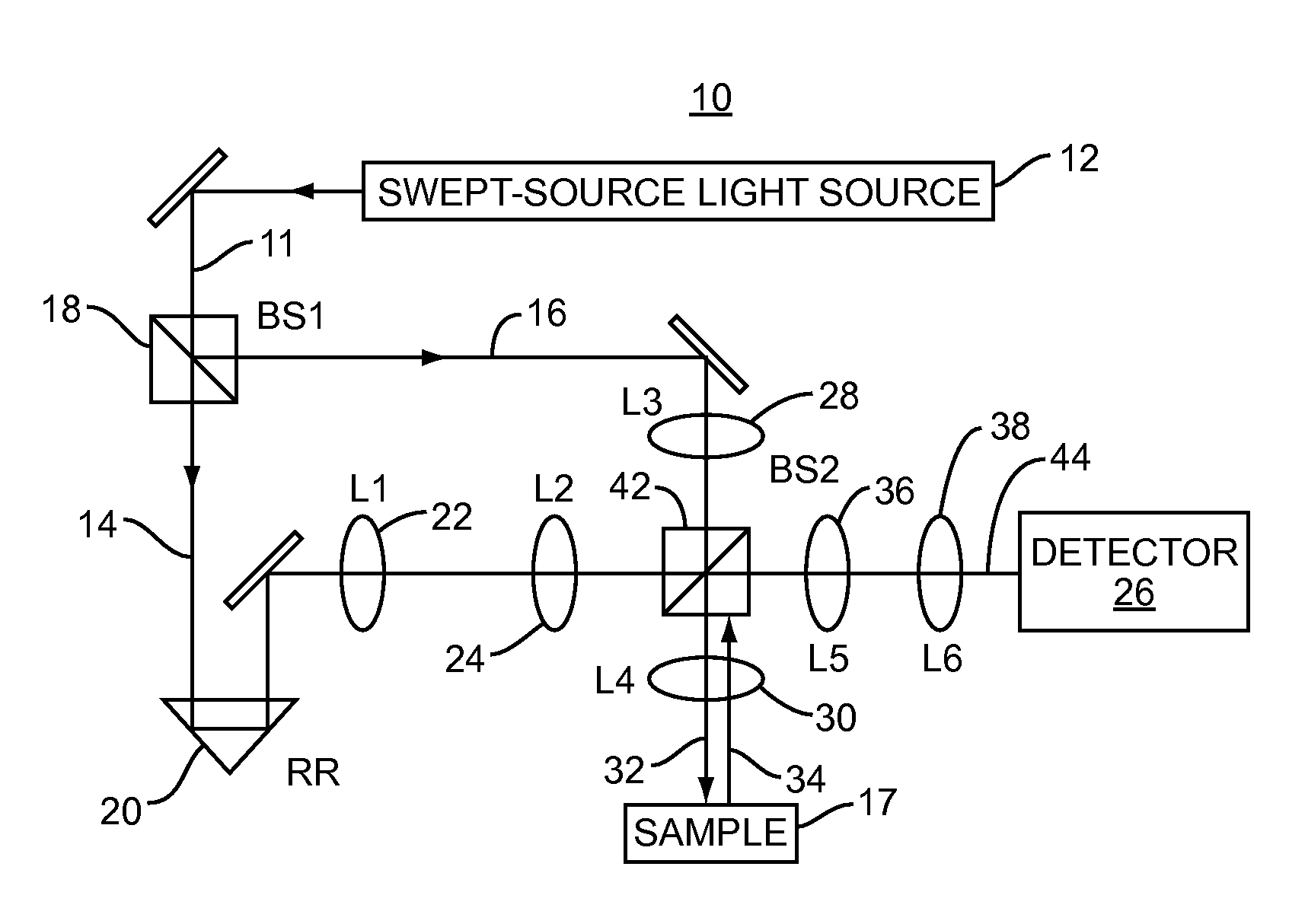
[0011]Embodiments disclosed herein involve low-coherence interferometry (LCI) techniques which enable acquisition of structural and depth information regarding a sample of interest at rapid rates. The acquisition rate is sufficiently rapid to make in vivo applications feasible. Biomedical applications of the embodiments disclosed herein include using the a / LCI systems and processes described herein for measuring cellular morphology in tissues and in vitro as well as diagnosing intraepithelial neoplasia, and assessing the efficacy of chemopreventive and chemotherapeutic agents. Prospectively grading tissue samples without tissue processing can also be accomplished using the embodiments disclosed herein, demonstrating the potential of the technique as a biomedical diagnostic.
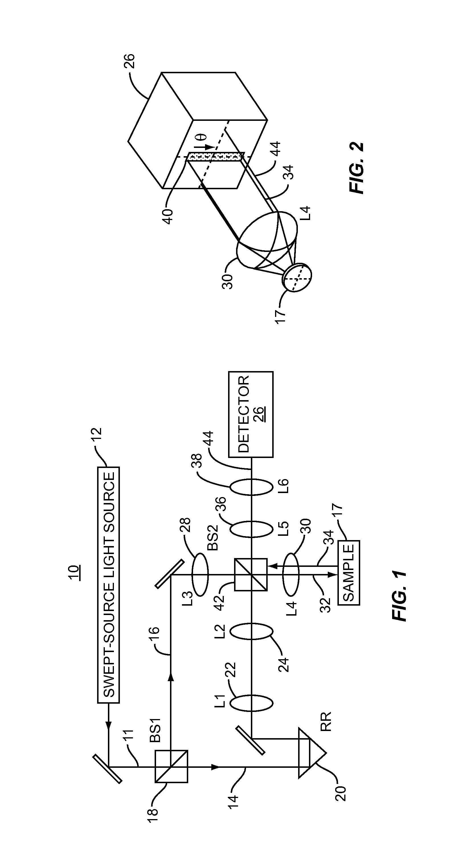
[0014]The spectrally-resolved and angle-resolved scattered information about the sample can be detected at a single scattering angle to provide a single scattering plane (i.e., 1-dimension) of spectrally-resolved and angle-resolved scattered information about the sample. Alternatively, the spectrally-resolved and angle-resolved scattered information about the sample can be detected at a plurality or range of angles to provide two-dimensional spectrally-resolved and angle-resolved scattered information about the sample. Capture of two-dimensional spectrally-resolved and angle-resolved scattered information from multiple scattering angles allows generation of more information about the sample under study and / or information with higher signal-to-noise ratio.
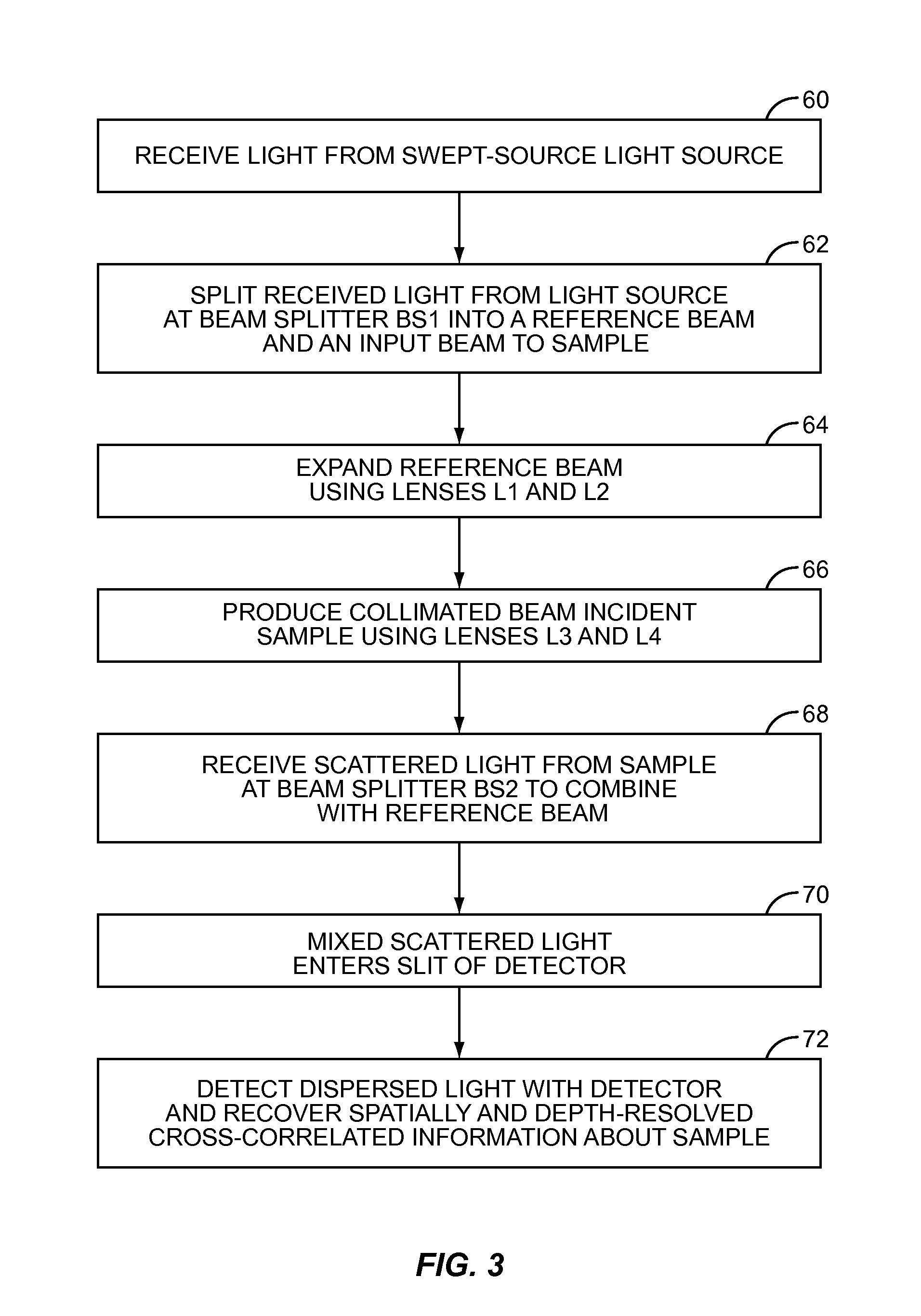
[0015]Depth information about the sample can be obtained using Fourier domain concepts as well as time domain techniques when using SS a / LCI. For example, in one manner of using time domain techniques to obtain depth information, the sample can be moved with respect to the light source to direct light at different planes within the sample. The resulting scattered light is processed to determine depth characteristics about the sample of interest. When using Fourier techniques as an example, the spectrally-resolved distribution of the scattered light returned from the sample as a result of the light emitted by the swept-source light source is converted into the Fourier domain. This allows obtaining depth-resolved information about the sample. Because the light source is swept, a spectrometer is not required to obtain spectral information about the sample, because the returned scattered light from the sample is already in the spectral domain as a result of a series of data acquisitions collected in narrower wavelengths or ranges emitted by the light source during its sweep. Scattering size characteristic information about the sample can be obtained by processing the spectrally-resolved and depth-resolved profile.
[0020]The LCI-based apparatuses, systems, and methods described above and in this application can be clinically viable methods for assessing tissue health without the need for tissue extraction via biopsy or subsequent histopathological evaluation. These LCI-based apparatuses, systems, and methods can be applied for a number of purposes including, but not limited to: early detection and screening for dysplastic tissues, disease staging, monitoring of therapeutic action, and guiding the clinician to biopsy sites. Some potential target tissues include the esophagus, the colon, the stomach, the oral cavity, the lungs, the bladder, and the cervix. The non-invasive, non-ionizing nature of the optical and LCI probe means that it can be applied frequently without adverse affect. The provision of rapid results through the use of the a / LCI systems and processes disclosed herein greatly enhance its widespread applicability for disease screening.

Problems solved by technology

Although microscopic examination has led to great advances in understanding cells and their structure, it is inherently limited by the artifacts of preparation.

Method used


Image

  • Apparatuses, systems, and methods for low-coherence interferometry (LCI)
  • Apparatuses, systems, and methods for low-coherence interferometry (LCI)
  • Apparatuses, systems, and methods for low-coherence interferometry (LCI)

Examples


Embodiment Construction

[0011]Embodiments disclosed herein involve low-coherence interferometry (LCI) techniques which enable acquisition of structural and depth information regarding a sample of interest at rapid rates. The acquisition rate is sufficiently rapid to make in vivo applications feasible. Biomedical applications of the embodiments disclosed herein include using the a / LCI systems and processes described herein for measuring cellular morphology in tissues and in vitro as well as diagnosing intraepithelial neoplasia, and assessing the efficacy of chemopreventive and chemotherapeutic agents. Prospectively grading tissue samples without tissue processing can also be accomplished using the embodiments disclosed herein, demonstrating the potential of the technique as a biomedical diagnostic.

[0012]In one embodiment, a “swept-source” (SS) light source is used in LCI to obtain structural and depth information about a sample. The swept-source light source is used to generate a reference signal and a sign...


PUM


Abstract

Embodiments described herein involve low-coherence interferometry (LCI) techniques which enable acquisition of structural and depth information regarding a sample of interest. In one embodiment, a “swept-source” (SS) light source is used in LCI to obtain structural and depth information about a sample. The swept-source light source can be used to generate a reference signal and a signal directed towards a sample. Light scattered from the sample is returned as a result and mixed with the reference signal to achieve interference and thus provide structural information regarding the sample. Depth information about the sample can be obtained using Fourier domain concepts as well as time domain techniques. Several LCI embodiments employing a swept-source light source are disclosed herein. In another embodiment disclosed herein, an a / LCI system and method is provided that is based on a time domain system and employs a broadband light source. The systems and processes disclosed herein can be used for biomedical applications, including measuring cellular morphology in tissues and in vitro as well as diagnosing intraepithelial neoplasia, and assessing the efficacy of chemopreventive and chemotherapeutic agents.

Description

RELATED APPLICATIONS[0001]This patent application claims priority to U.S. Provisional Patent Application Ser. No. 60 / 971,980, filed on Sep. 13, 2007 and entitled “Systems and Methods for Angle-Resolved Low Coherence Interferometry,” which is incorporated herein by reference in its entirety.BACKGROUND[0002]1. Field of the Invention[0003]The technology of the present application relates generally to low-coherence interferometry (LCI) and obtaining structural and depth-resolved information about a sample using LCI. The technology includes angle-resolved-based LCI (a / LCI), Fourier-based LCI (f / LCI), and Fourier and angle-resolved-based LCI (fa / LCI) apparatuses, systems, and methods.[0004]2. Technical Background[0005]Examining the structural features of cells is essential for many clinical and laboratory studies. The most common tool used during examination for the study of cells has been the microscope. Although microscopic examination has led to great advances in understanding cells an...

Claims


Application Information

Patent Timeline
IPC IPC(8): G01B9/02
CPCA61B5/0059G01N21/4795G01B9/02004G01B9/02088G01B9/0209G01B9/02043G01B9/02084G01B9/02044
Inventor WAX, ADAMBROWN, WILLIAM J.
Owner DUKE UNIV
Learn More