Ultrasound image diagnosis apparatus

一种图像诊断、超声波的技术,应用在声波诊断、次声波诊断、超声波/声波/次声波诊断等方向,能够解决主要部分不容易看清、视认性、操作性差、视认性、操作性变差等问题,达到提高视认性、减轻负担、交换容易的效果

Active Publication Date: 2010-11-17
TOSHIBA MEDICAL SYST CORP
View PDF4 Cites 7 Cited by
  • Summary
  • Abstract
  • Description
  • Claims
  • Application Information

AI Technical Summary

Problems solved by technology

However, in order to easily see the monitor screen of the ultrasonic diagnostic imaging device, the indoor lighting of the examination room during the ultrasonic examination is very dark.
Therefore, the removal and reinstallation of the connector to replace the ultrasonic probe has the difficulty of deteriorating visibility and operability.
[0007] In addition, the main part of the main body of the ultrasonic diagnostic imaging device other than the operation panel is not easy to see, so the visibility and operability such as confirmation of examination records are also poor.

Method used


Image

  • Ultrasound image diagnosis apparatus
  • Ultrasound image diagnosis apparatus
  • Ultrasound image diagnosis apparatus

Examples


Embodiment Construction

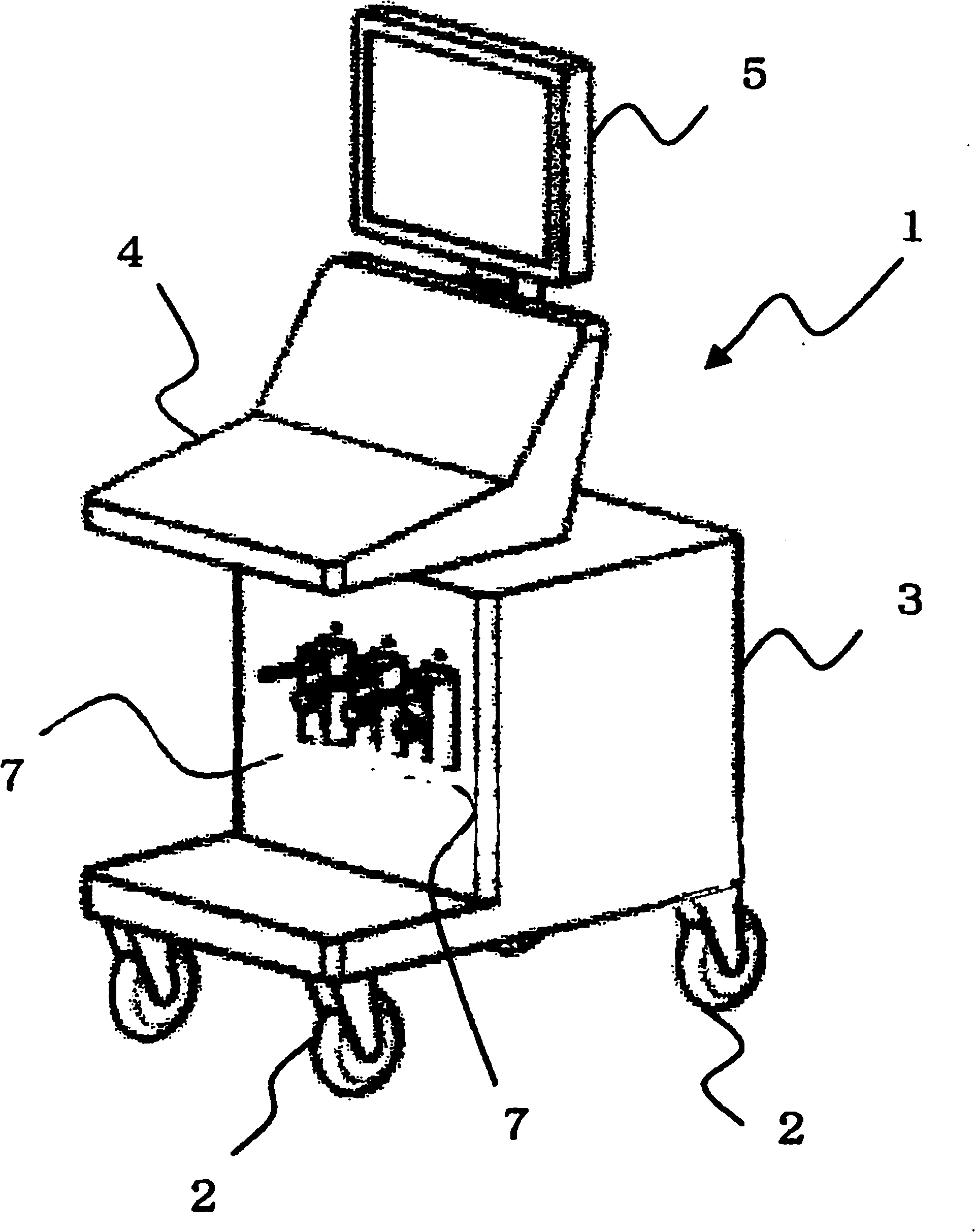
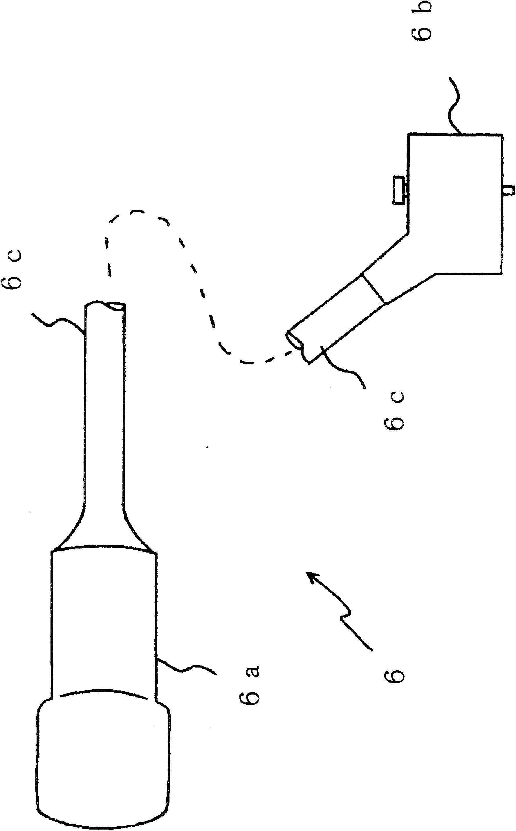
[0016] Such as figure 1 As shown, an ultrasonic imaging diagnostic apparatus 1 includes an ultrasonic imaging diagnostic apparatus main body 3 , an operation panel 4 , a display 5 for displaying ultrasonic images, and an ultrasonic probe 6 for transmitting and receiving ultrasonic signals to a subject. The operation panel 4 has a touch panel and a keyboard for the operator to perform various operations, and is usually provided on the upper part of the main body 3 of the ultrasonic imaging diagnostic apparatus. Casters 2 are attached in order to move the ultrasonic imaging diagnostic apparatus main body 3 easily.

[0017] A connector 7 for electrically connecting the ultrasonic probe 6 is attached to the main body 3 of the ultrasonic imaging diagnostic apparatus. For example, as shown in the figure, the connector is arranged on the front side of the main body 3 of the ultrasonic imaging diagnostic apparatus, and is located below the operation panel 4 . In the present embodime...


PUM


Abstract

An ultrasound image diagnosis apparatus executing lighting-on and lighting-off of a lighting device by detecting an identification (ID) data of a probe or an operation on an operation panel. The ultrasound image diagnosis also includes a lighting device configured to identify a usage state or non-usage state of the ultrasound probe by a lighting color. While an ultrasound examination is performedin a dimly-lit room, a visibility of an ultrasound probe connector can be increased.

Description

[0001] This application claims priority under 35 U.S.C §119(a) to Japanese Patent Application No. 2009-115872 filed May 12, 2008, the entire contents of which are incorporated herein by reference. technical field [0002] The present invention relates to an ultrasonic imaging diagnostic apparatus, and in particular, to an ultrasonic imaging diagnostic apparatus that can easily perform various tasks such as maneuvering an ultrasonic probe in a dark examination room. Background technique [0003] Ultrasonic imaging diagnostic device is a device that sends ultrasonic waves to the subject through an ultrasonic probe, receives echo signals from the subject, and reproduces ultrasonic images based on the echo signals, and inspects the organs and blood flow of the subject. , are widely used in the medical field. When performing ultrasonic image diagnosis, appropriate ultrasonic probes with different frequencies and vibrator surface shapes are used for abdominal use, circulatory syst...

Claims


Application Information

Patent Timeline
Patent Type & Authority Applications(China)
IPC IPC(8): A61B6/00
CPCA61B8/00A61B8/4405A61B8/4438
Inventor 佐藤友广平久井克也仓俣胜辉小野寺英雄
Owner TOSHIBA MEDICAL SYST CORP
Why Eureka
Try Eureka