Suspected lung nodule image enhancement directional scale filtering method

An image enhancement and image technology, applied in image enhancement, image data processing, instruments, etc., can solve the problem of image enhancement of suspected lung nodes, and achieve the effect of improving visual effect and gray contrast enhancement.

Inactive Publication Date: 2011-03-23
XIAN UNIV OF TECH
View PDF1 Cites 11 Cited by
  • Summary
  • Abstract
  • Description
  • Claims
  • Application Information

AI Technical Summary

Problems solved by technology

[0010] The purpose of the present invention is to provide a method for enhancing direction-scale filtering of suspected pulmonary nodule images, which solves the problem that it is difficult to enhance suspected pulmonary nodule images in chest X-rays in the prior art

Method used


Image

  • Suspected lung nodule image enhancement directional scale filtering method
  • Suspected lung nodule image enhancement directional scale filtering method
  • Suspected lung nodule image enhancement directional scale filtering method

Examples


Embodiment Construction

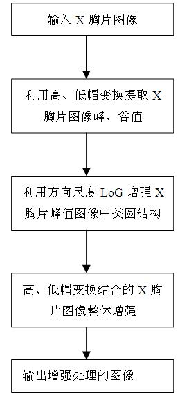
[0045] The present invention will be described in detail below in conjunction with the accompanying drawings and specific embodiments.

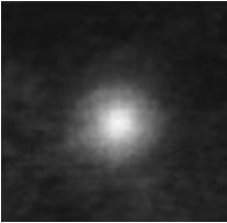
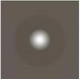
[0046] Clinical Observation [13] It shows that the shape of a typical lung node is usually approximated as a sphere, and its density is equivalent to that of water, which is slightly larger than the lung fluid around it. Therefore, on the X-ray chest film, the lung nodes generally appear as brighter interiors of different sizes The outer darker circle (as in figure 1 shown)

[0047] The Laplacian Gaussian function can be described mathematically as:

[0048] Formula 1)

[0049] In the formula, It is the standard deviation of the Gaussian function, which is used to control the scale of the filter; x and y represent the gray value of the pixel in the horizontal and vertical directions, respectively; e represents the base of the natural logarithm; π is the pi. The function is an isotropic circular distribution function [14] . when ...


PUM


Abstract

The invention discloses a suspected lung nodule image enhancement directional scale filtering method, which comprises the following steps: inputting an original image, and calculating a peak value image and a valley value image of the original X-ray chest radiography respectively by utilizing a top-hat transform operator and a bottom-hat transform operator in gray morphology; carrying out directional enhancement on a lung similar-round focus image in the X-ray chest peak value image by utilizing a directional scale Laplace Gauss function as a matched filter, introducing a visual correction factor in image enhancement operation, and carrying out visual correction on a convolution image of a directional scale Laplace Gauss operator; and adding the original X-ray chest image in the image obtained from the step 2, and then subtracting the valley value image obtained from the step 1 to obtain an output image. The beneficiary effect of the invention is that the grey contrast between the lung similar-round structural focus in the X-ray chest image and the periphery background tissue of the lung similar-round structural focus is enhanced obviously, the texture details in the X-ray chest are maintained, and the visual effect of the X-ray chest is improved preferably.

Description

technical field [0001] The invention belongs to the technical field of automatic analysis and processing of medical images, and in particular relates to an image enhancement direction scale filtering method for suspected pulmonary nodules based on mathematical morphology improvement. Background technique [0002] The mortality rate of lung cancer ranks first in the number of deaths caused by malignant tumors in the world. The 5-year survival rate of patients is closely related to the tumor stage. The overall 5-year survival rate of lung cancer patients is about 15%. However, the 5-year survival rate of patients with stage I up to 70% [1] Therefore, early detection and early diagnosis are of great significance to improve the survival rate of lung cancer patients. [0003] Lung cancer is manifested in the form of lung nodes in the early stage. Helical Computed Tomography Scans (HCTS) and Chest Radiography (Chest Radiography) are the two main technical means for medical image...

Claims


Application Information

Patent Timeline
Patent Type & Authority Applications(China)
IPC IPC(8): G06T5/00
Inventor 石争浩白军
Owner XIAN UNIV OF TECH
Why Eureka
Try Eureka