
3-D ultrasound imaging

An ultrasonic imaging system and ultrasonic scanning technology, applied in ultrasonic/sonic/infrasonic diagnosis, sonic diagnosis, infrasonic diagnosis, etc., can solve problems such as time-consuming, high skill level, and error-prone

Inactive Publication Date: 2011-09-21
KONINK PHILIPS ELECTRONICS NV
View PDF4 Cites 10 Cited by
  • Summary
  • Abstract
  • Description
  • Claims
  • Application Information

AI Technical Summary

Problems solved by technology

However, finding this standard view that allows for a reliable diagnosis still requires a relatively high level of skill
Furthermore, this process is relatively error-prone and time-consuming

Method used


Image

  • 3-D ultrasound imaging
  • 3-D ultrasound imaging
  • 3-D ultrasound imaging

Examples


Embodiment Construction

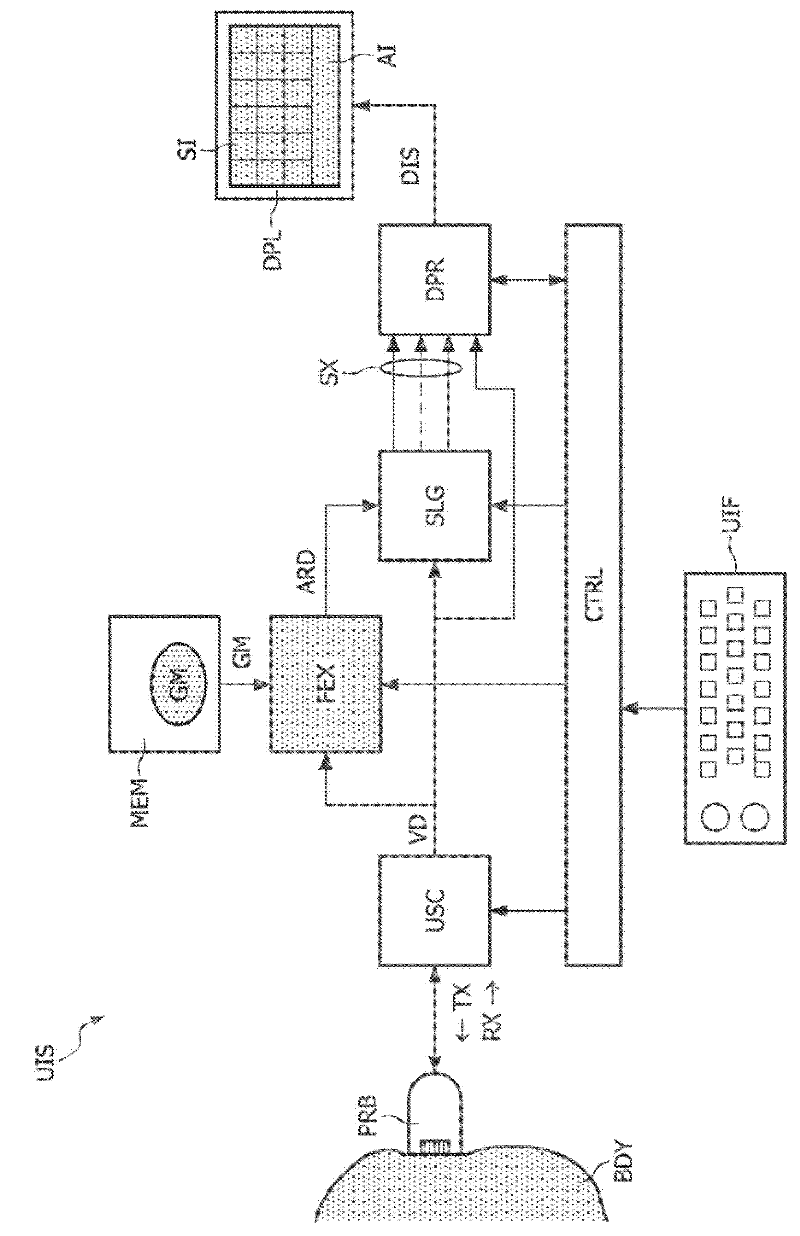
[0024] figure 1 An ultrasound imaging system UIS capable of performing 3-D ultrasound scans is illustrated. The ultrasound imaging system UIS comprises various functional entities making up the ultrasound imaging acquisition-and-processing path: probe PRB, ultrasound scanning component USC, feature extractor FEX, slice generator SLG, and display processor DPR. The probe PRB may for example comprise a two-dimensional array of piezoelectric transducers. The ultrasound scanning component USC may include an ultrasound transmitter and an ultrasound receiver, which may each contain a beamforming module. The ultrasound scanning component USC may further comprise one or more filter modules, a so-called B-mode processing module, and a Doppler-mode processing module.

[0025] The feature extractor FEX can be implemented by means of a set of instructions already loaded into a programmable processor, for example. In this software-based implementation, the set of instructions defines th...


PUM


Abstract

In an ultrasound imaging system (UIS), an ultrasound scanning assembly (USC) provides volume data (VD) resulting from a three-dimensional scan of a body (BDY). A feature extractor (FEX) searches for a best match between the volume data (VD) and a geometrical model (GM) of an anatomical entity. The geometrical model (GM) comprises respective segments representing respective anatomic features. Accordingly, the feature extractor (FEX) provides an anatomy-related description (ARD) of the volume data (VD), which identifies respective geometrical locations of respective anatomic features in the volume data (VD). In a preferred embodiment, a slice generator (SLG) generates slices (SX) from the volume data (VD) based on the anatomy-related description (ARD) of the volume data (VD).

Description

technical field [0001] One aspect of the invention relates to an ultrasound imaging system capable of performing three-dimensional (3-D) ultrasound scans to generate volume data. The ultrasound imaging system may be useful, for example, in fetal examination, especially in the detection of structural abnormalities in the fetal heart. Other aspects of the invention relate to ultrasound imaging methods, and computer program products. Background technique [0002] A 3-D ultrasound scan typically involves transmitting ultrasound waves that so to speak illuminate a specific volume within the body that can be designated as a target volume. This can be done, for example, by emitting ultrasound at several different angles. Volumetric data is obtained by receiving and processing reflected waves. The volume data is representative of the target volume within the body. The volumetric data can be displayed on a display device in a manner that provides a three-dimensional representatio...

Claims


Application Information

Patent Timeline
Patent Type & Authority Applications(China)
IPC IPC(8): G01S15/89
CPCA61B8/5238A61B8/0883G01S15/8977G01S15/8993A61B8/523A61B8/483A61B8/0866A61B8/0833
Inventor A·科莱-比永J·杰戈A·道L·庞弗里
Owner KONINK PHILIPS ELECTRONICS NV
Learn More