Coccidia cell culture model and application thereof

A cell culture and cell technology, applied in the direction of embryonic cells, animal cells, vertebrate cells, etc., to achieve the effect of strong invasion ability

Inactive Publication Date: 2011-10-19
SHANGHAI VETERINARY RES INST CHINESE ACAD OF AGRI SCI
View PDF0 Cites 0 Cited by
  • Summary
  • Abstract
  • Description
  • Claims
  • Application Information

AI Technical Summary

Problems solved by technology

So far, there have been no public reports on the application of the DF-1 cell culture

Method used


Image

  • Coccidia cell culture model and application thereof
  • Coccidia cell culture model and application thereof
  • Coccidia cell culture model and application thereof

Examples


Embodiment 1

[0035] Example 1 Collection and purification of Eimeria tenella sporozoites

[0036] The purification of oocysts refers to the separation and purification technology of coccidia oocysts in the "Advanced Parasitology Experiment Guide", with slight modifications during the experimental operation (Suo Xun et al., 2006).

[0037] (1) The sporulated oocysts of Eimeria tenella were pressed by 1×10 4 Inoculate 14-day-old coccidial-free chicks per piece / only. On the 8th day after infection, the chickens were dissected and the cecum was collected to collect unsporulated oocysts. They were cultured at 28°C until they were fully sporulated, and the sporulated oocysts were collected and stored at 4°C. The period is 1-2 months.

[0038] (2) The sporulated oocysts of E.tenella stored at 4°C were centrifuged at 2500r / min for 10min at room temperature. After removing potassium dichromate, add an appropriate amount of distilled water to the precipitate, suspend the precipitate, centrifuge at...

Embodiment 2

[0045] The establishment of embodiment 2 Eimeria tenella DF-1 cell culture model

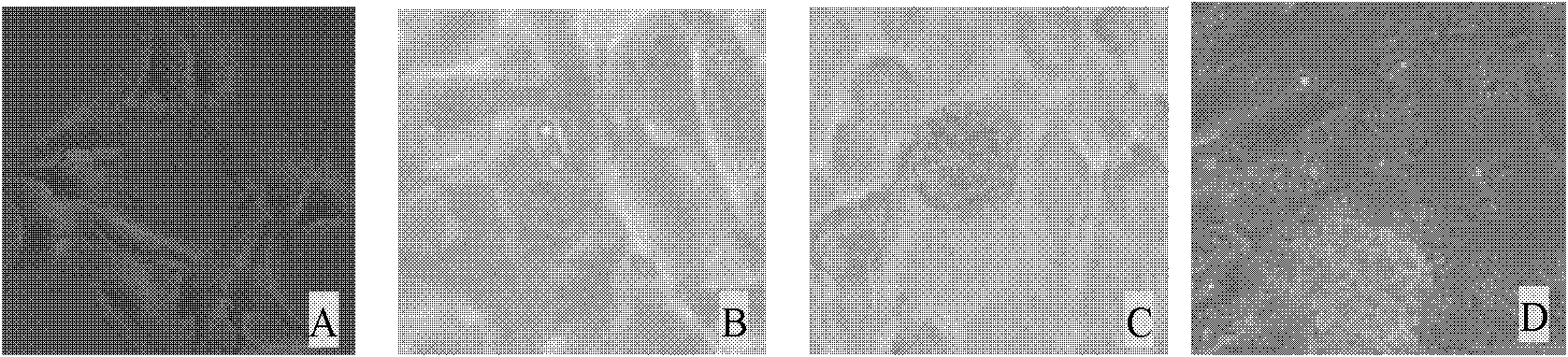
[0046] Digest well-growing DF-1 cells with 0.25% trypsin, collect the cells after 2-4 minutes, count with a cell counting plate, dilute with 10% DMEM, and dilute at 1×10 5 Spread 24-well plates at 37°C, 5% CO 2 Cultured for 24h (see figure 1 A), each hole access 1 × 10 5 Sporozoites, 41°C, 5% CO 2 Culture, it can be seen that sporozoites can invade DF-1 cells within 24h (such as figure 1 B), developed into the 1st generation schizont within 60h (see figure 1 C), at 72h, a large number of free first-generation merozoites can be observed in the cell supernatant (see figure 1 D), a small number of merozoites can continue to invade DF-1 cells, but no merozoites continue to develop into oocysts.

Embodiment 3

[0047] Example 3 Application of the Eimeria tenella DF-1 cell model in establishing a detection method for sporozoite invasion activity

[0048] 1. Establishment of detection method for invasion activity of Eimeria tenella sporozoites

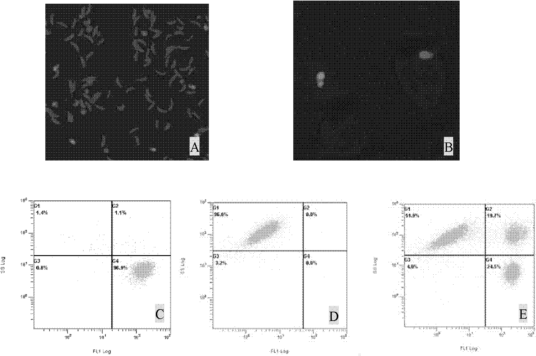
[0049] Count the purified sporozoites of Eimeria tenella, label the sporozoites according to the CFDA SE cell proliferation and tracer detection kit operation steps, inoculate DF-1 cell culture, wash twice with PBS before detection, trypsinize and collect Cells, detect the invasion cell number of coccidia sporozoites with flow cytometer, thereby calculate the invasion rate of sporozoites, the formula is: the invasion rate of sporozoites=100%×[invasion cell number / (non-invasion cell number+invasion cell number)]. Normal DF-1 cells and sporozoites labeled with CFDA SE (fluorescent probe 5(6)-carboxyfluorescein diacetate succinimidyl ester) were used as control groups. It can be seen that the fluorescence of sporozoites labeled with CFDA SE is v...


PUM


Abstract

The invention discloses a coccidia cell culture model. The coccidia cell culture model is built through the processes that sporozoites of Eimeria tenella are inoculated into DF-1 cells of a Chicken Embryo Fibroblast Cell line and grow into first generation merozoites. The invention also discloses an application of the coccidia cell culture model in a selection of anticoccidial drugs. The coccidia cell culture model can be utilized for establishment of a test method for measuring objectively sporozoite invasion activity, and is suitable for a selection of anticoccidial drugs and researches of gene or protein functions during a coccidia growth process.

Description

technical field [0001] The invention relates to a cell culture model, in particular to a coccidian cell culture model and its application. Background technique [0002] Chicken coccidiosis is a protozoan disease caused by Eimeria coccidiosis parasitizing on chicken intestinal epithelial cells. It is widely distributed all over the world and is one of the important diseases that endanger the chicken industry. Its life history is relatively complicated, and it needs to go through developmental stages such as sporulation, schizogenesis, and gametogenesis. Cell culture can provide a clean and pollution-free environment for coccidia research, and at the same time make people eager to study the mechanism of action and activity of anticoccidial drugs and the development, behavior, structure, and immunity of coccidia in an "uncontrolled" and "transparent" environment. , heredity, cytochemistry and biochemistry, etc. dreams become reality. The subculture of coccidia is a process in...

Claims


Application Information

Patent Timeline
IPC IPC(8): C12N5/073C12Q1/02G01N15/10
Inventor 姜连连黄兵韩红玉董辉赵其平朱顺海马卫娇曾艳波程军李婷孔春林
Owner SHANGHAI VETERINARY RES INST CHINESE ACAD OF AGRI SCI
Why Eureka
Try Eureka