Three-dimensional lung vessel image segmentation method based on geometric deformation model

A deformation model and blood vessel image technology, applied in image analysis, image data processing, 3D modeling, etc., can solve problems such as speed and accuracy that cannot meet application requirements

Inactive Publication Date: 2011-11-16
NORTHEASTERN UNIV
View PDF5 Cites 26 Cited by
  • Summary
  • Abstract
  • Description
  • Claims
  • Application Information

AI Technical Summary

Problems solved by technology

[0007] In view of the problems that the speed and accuracy of the pulmonary vessel segmentation method used in the prior art cannot meet the application requirements, the te

Method used


Image

  • Three-dimensional lung vessel image segmentation method based on geometric deformation model
  • Three-dimensional lung vessel image segmentation method based on geometric deformation model
  • Three-dimensional lung vessel image segmentation method based on geometric deformation model

Examples


Embodiment Construction

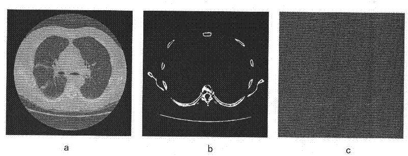
[0043] The practice of the present invention utilizes multi-slice helical CT (HRCT) image data. Because CT images can provide high-definition images and provide high contrast for various tissues in the images, they are usually used in the diagnosis of lung diseases. With the development of multi-slice spiral CT, doctors can obtain higher-resolution images, minimize the local volume effect, and obtain more patient information through one detection, further expanding the application of CT images.

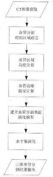
[0044] Combined with the accompanying drawings, the flow chart of the three-dimensional pulmonary vessel image segmentation based on the geometric deformation model is as follows: figure 1 As shown, the detailed segmentation method of the present invention comprises the following five steps:

[0045] (1) The initial segmentation area is determined;

[0046] (2) Calculation of the average value of the blood vessel area;

[0047] (3) Calculation of blood vessel edge energy;

[0048]...


PUM


Abstract

The invention provides a three-dimensional lung vessel image segmentation method based on a geometric deformation model. The method comprises the following steps: (1) determining vessel segmentation computing regions according to the physiological structure characteristics of a human body, wherein region selection completely covers targets to be segmented and the shape characteristics of the regions are stable, thereby avoiding computing a global region and improving segmentation speed; (2) computing the mean value of the vessel regions and positioning internal and external homogeneous regions of the targets; (3) computing vessel edge energy and evolving a curved surface along second derivatives in an image gradient direction so that the curved surface is accurately converged to a target edge; (4) correspondingly establishing a three-dimensional vessel segmentation curved surface evolution model and effectively combining the mean value and edge energy of the internal and external regions of the lung vessels; and (5) adopting optimized level set evolution for obtaining solution according to the established deformation model and impliedly solving a curved surface motion according to the level set function curved surface evolution. A large quantity of lung CT image experiments proof that the method provided by the invention has the advantages of rapid and accurate lung vessel segmentation and strong robustness.

Description

technical field [0001] The invention belongs to the intersection field of digital image processing and medical imaging technology, and particularly relates to a three-dimensional pulmonary blood vessel image segmentation method based on a geometric deformation model. Background technique [0002] Because CT equipment can provide high-definition images and provide high contrast for various human tissues in the images, it is usually used in the diagnosis of lung diseases. Pulmonary blood vessels are one of the most important tissues and organs with the most complex topological structure in the human body. The precise segmentation of pulmonary vessels is the key to the detection of lung cancer lesions. This is because the pulmonary vessels are mostly distributed near the lung cancer lesions in CT images, and the gray values ​​of the two are similar, which is an important interference in the detection of lung cancer lesions. In addition, pulmonary vessel segmentation is also of...

Claims


Application Information

Patent Timeline
IPC IPC(8): G06T7/00G06T17/00
Inventor 贾同魏颖吴成东
Owner NORTHEASTERN UNIV
Why Eureka
Try Eureka