Ultrasonic imaging system for elasticity measurement and method for measuring elasticity of biological tissue

An ultrasonic imaging system and ultrasonic imaging technology, applied in the field of ultrasonic imaging systems for measuring the elasticity of biological tissue, can solve the inconvenience of obtaining information on the elasticity of biological tissue, the difficulty of realization, and the complexity of the ultrasonic elastography device or method, etc. problems, to achieve the effect of improving convenience and accuracy, improving accuracy, and reducing the difficulty of processing

Active Publication Date: 2014-05-21
LEPU MEDICAL TECH (BEIJING) CO LTD
View PDF4 Cites 0 Cited by
  • Summary
  • Abstract
  • Description
  • Claims
  • Application Information

AI Technical Summary

Problems solved by technology

However, both the device and the method of ultrasonic elastography are relatively complicated, and the implementation is relatively difficult, which is very inconvenient in the process of obtaining elastic information of biological tissues.

Method used


Image

  • Ultrasonic imaging system for elasticity measurement and method for measuring elasticity of biological tissue
  • Ultrasonic imaging system for elasticity measurement and method for measuring elasticity of biological tissue
  • Ultrasonic imaging system for elasticity measurement and method for measuring elasticity of biological tissue

Examples


Embodiment Construction

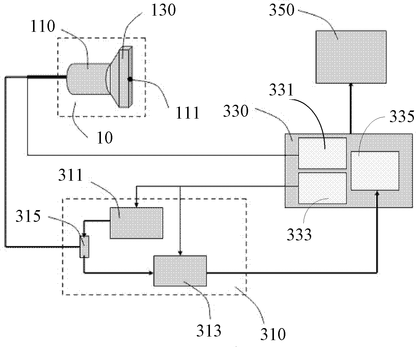
[0048] figure 1 An ultrasonic imaging system for elasticity measurement in an embodiment is shown, the system includes a probe 10 , an ultrasonic imaging device 310 , a control and processing device 330 and a display device 350 .

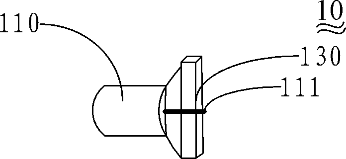
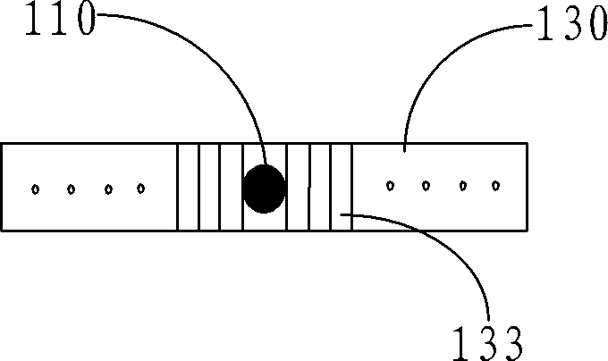
[0049] The probe 10 includes a low-frequency oscillation driving device 110 and an ultrasonic transducer array 130 .

[0050] The low-frequency oscillation driving device 110 is used to generate vibration and form a shear wave propagating from the body surface to the inside of the tissue.

[0051] In this embodiment, the low-frequency oscillation driving device 110 is a low-frequency oscillator or a motor. In order to cause micro-deformation of biological tissue by external force or internal force, the vibration shaft 111 in the low-frequency oscillating driver 110 vibrates at low frequency and low amplitude, causing shear waves propagating into the biological tissue and inducing micro-deformation.

[0052]If the frequency of the shear wave in the...


PUM


Abstract

An ultrasonic imaging system for measuring elasticity of biological tissues, including a probe (10), an ultrasonic imaging device (310), a controlling and processing device (330), and a display device (350). The probe(10) includes a low frequency oscillating device (110) and an ultrasonic array transducer (130). The low frequency oscillating device (110) is configured for generating oscillation and forming shear waves propagating from a body surface to the biological tissues. The ultrasonic array transducer (130) is configured for emitting ultrasonic signals to the biological tissues and receiving ultrasonic echo signals reflected from the biological tissues. The ultrasonic imaging device (310), according to loaded imaging parameters, drives the ultrasonic array transducer (130) to emit the ultrasonic signal to the biological tissues, and receives and processes the ultrasonic echo signals from the ultrasonic array transducer (130). The controlling and processing device (330) controls the low frequency oscillating device (110) and the ultrasonic imaging device (310) to obtain a 2D ultrasonic image and elasticity information of the biological tissues. The display device (350) displays the 2D ultrasonic image and the elasticity information of the biological tissues. The 2D ultrasonic image and elasticity information of the biological tissues can be obtained at the same time, such that accuracy of clinical diagnosing is promoted. The low frequency oscillating device (110) and the ultrasonic array transducer (130) are independent, so the ultrasonic array transducer (130) will not move with the low frequency oscillating device (110), and there is no need to considerate displacement compensation. The probe (10) can locate accurately with the aid of the 2D ultrasonic image, such that convenience and accuracy is promoted in measuring elastic modulus and process difficulty is decreased.

Description

【Technical field】 [0001] The invention relates to ultrasonic imaging technology, in particular to an ultrasonic imaging system for measuring elasticity and a method for measuring elasticity of biological tissue. 【Background technique】 [0002] Traditional B-mode ultrasound imaging is mainly used to obtain physiological structure information of biological tissues, and is not sensitive to early lesions, but the mechanical characteristics of biological tissues have changed significantly during early lesions. The mechanical characteristics such as elastic modulus (hardness) of biological tissues depend on the molecular composition and corresponding microstructure of biological tissues, and are closely related to their physiological and pathological characteristics. Diseased tissues and normal tissues often have mechanical characteristics such as elastic modulus (hardness) Therefore, the detection of changes in the mechanical characteristics of biological tissues can provide an i...

Claims


Application Information

Patent Timeline
Patent Type & Authority Patents(China)
IPC IPC(8): A61B8/08
CPCG01S7/52042A61B8/485A61B8/085A61B8/0825
Inventor 郑海荣凌涛李彦明姚慧曾成志
Owner LEPU MEDICAL TECH (BEIJING) CO LTD
Why Eureka
Try Eureka