
A radiological image stitching device and method

A splicing device and image technology, which is applied in the fields of radiological diagnosis, medical science, diagnosis, etc., can solve the problems of inconvenient shooting of the end of the foot, long one-way movement of the machine, and long time interval between two splicing, etc. The effect of increasing the shooting range and reducing the time

Active Publication Date: 2015-09-23
SHENZHEN MINDRAY BIO MEDICAL ELECTRONICS CO LTD
View PDF8 Cites 0 Cited by
  • Summary
  • Abstract
  • Description
  • Claims
  • Application Information

AI Technical Summary

Problems solved by technology

[0008] The existing scheme has the following defects: the one-way movement of the machine is long, resulting in a long time interval between the two stitchings; for standing filming, because the height of the detector has a lower limit, it is inconvenient for shooting the end of the foot (often requires a foot Pad); when filming in the supine position, due to the influence of the horizontal and lateral movement of the detector under the examination bed, sometimes it is impossible to achieve the shooting of the complete splicing part

Method used


Image

  • A radiological image stitching device and method
  • A radiological image stitching device and method
  • A radiological image stitching device and method

Examples


Embodiment Construction

[0025] The present invention will be further described in detail below through specific embodiments in conjunction with the accompanying drawings.

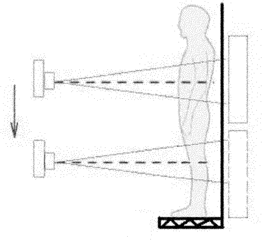
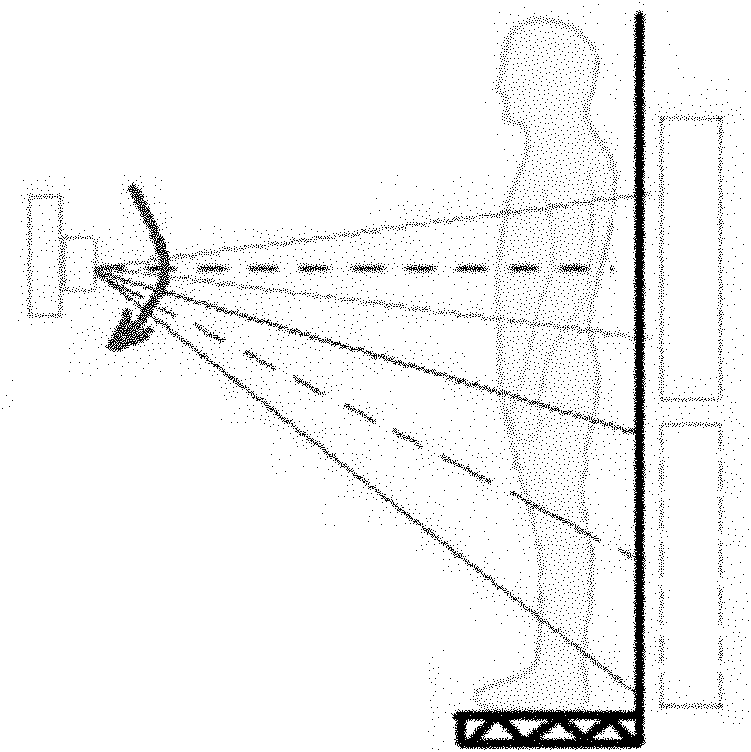
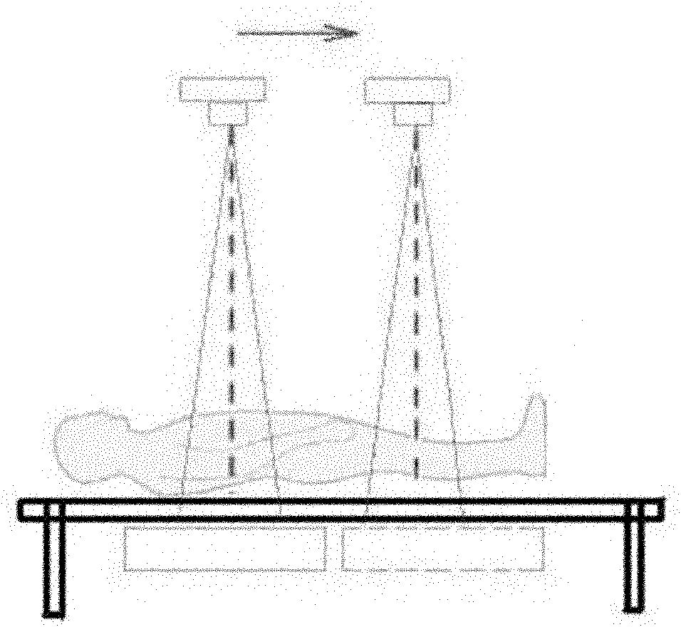
[0026] The radiation image splicing device disclosed herein includes a machine head and a detector, a motion control unit for controlling the movement of the machine head and the detector, and a supporting motion device for carrying imaging targets. The motion control unit performs splicing on the machine head During the photographing process, the relative movement between the supporting motion device and the detector is controlled.

[0027] In embodiment one: as Figure 5 As shown, for upright stitching, the radiographic image stitching device includes a supporting motion device, which is a lifting and lowering splicing bracket. During the splicing shooting process, in addition to the top-down movement of the machine head and the detector, the motion control unit also controls the bottom-up movement of the elevating splicing bra...


PUM


Abstract

The invention discloses a radiograph mosaic device, which comprises a head, a detector, a movement control unit and a supporting movement device. The movement control unit controls movement of the head and movement of the detector, the supporting movement device is used for carrying people, and the movement control unit controls the supporting movement device and the detector to move relatively in a mosaic photographing process of the head. As the movement control unit controls the supporting movement device and the detector to move relatively in the mosaic photographing process of the head, the radiograph mosaic device has the advantages that the image shooting and mosaic time is shortened, and a shooting range is expanded.

Description

technical field [0001] The present invention relates to an image processing method and device, in particular to an image splicing method and device for digital X-ray imaging (Digitalradiography, DR for short) equipment. Background technique [0002] Usually, in DR photographic inspection, the spine and legs of a person cannot be fully displayed in one exposure area. Through multiple exposures in sub-regions, and then stitching the pictures into one through image processing means, it is feasible to form full-frame image data. The current DR image stitching function uses the method of patient stillness, detector and machine head movement to complete the exposure action of each part. The head is the abbreviation of the X-ray tube frame, which is the output end mechanism of the X-ray, and is mainly connected by the ball tube and the speed limiter. [0003] There are roughly four types: [0004] Standing position and parallel movement: the detector is vertical, and the machine...

Claims


Application Information

Patent Timeline
Patent Type & Authority Patents(China)
IPC IPC(8): A61B6/00
Inventor 刘兵全张继晔文琪
Owner SHENZHEN MINDRAY BIO MEDICAL ELECTRONICS CO LTD
Learn More