
Data acquisition device of three-dimensional ultrasound image based on rear-end scanning

A data acquisition, three-dimensional ultrasound technology, applied in ultrasonic/sonic/infrasonic diagnosis, sonic diagnosis, infrasonic diagnosis and other directions, can solve problems such as insufficient accuracy, small scanning angle, complicated operation, etc. Improvement in the quality of results, the effect of easy operation

Active Publication Date: 2012-07-25
维视医学影像有限公司
View PDF4 Cites 12 Cited by
  • Summary
  • Abstract
  • Description
  • Claims
  • Application Information

AI Technical Summary

Problems solved by technology

Sector scan and scan are the most frequently used scanning methods, but the traditional sector scan and scan have inevitable defects: the anatomical structure of the chest organs cannot be scanned due to the influence of the ribs
[0004] This scanning mode successfully avoids the influence of the ribs of the human body, but the scanning device has the disadvantages of being complicated and heavy, complicated operation, small scanning angle, and insufficient precision.

Method used


Image

  • Data acquisition device of three-dimensional ultrasound image based on rear-end scanning
  • Data acquisition device of three-dimensional ultrasound image based on rear-end scanning
  • Data acquisition device of three-dimensional ultrasound image based on rear-end scanning

Examples


Embodiment Construction

[0026] The present invention will be described in further detail below in conjunction with the accompanying drawings and embodiments.

[0027] The mechanical device of the present invention can be realized by processing any material that meets the requirements, and the material used in this embodiment is aluminum.

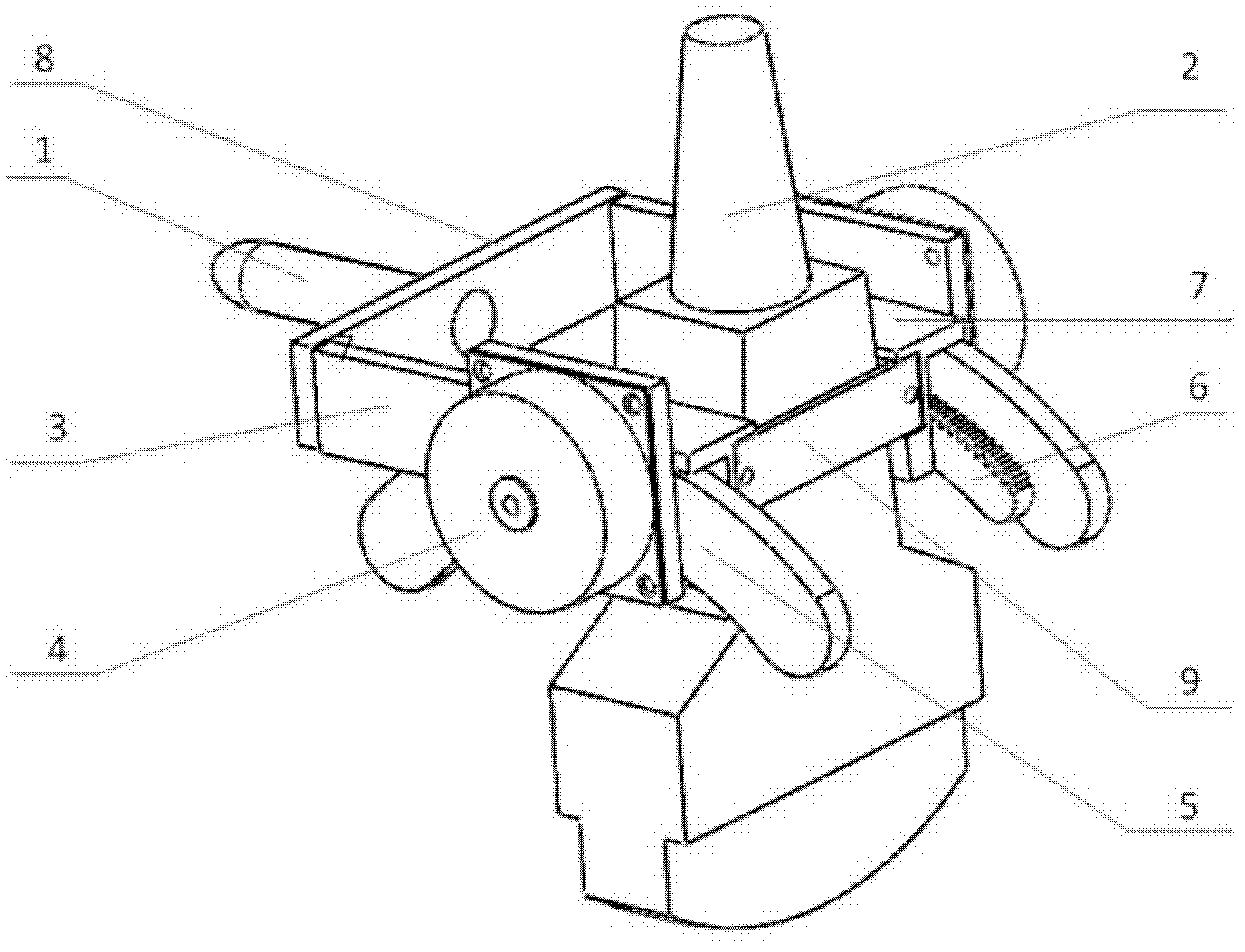
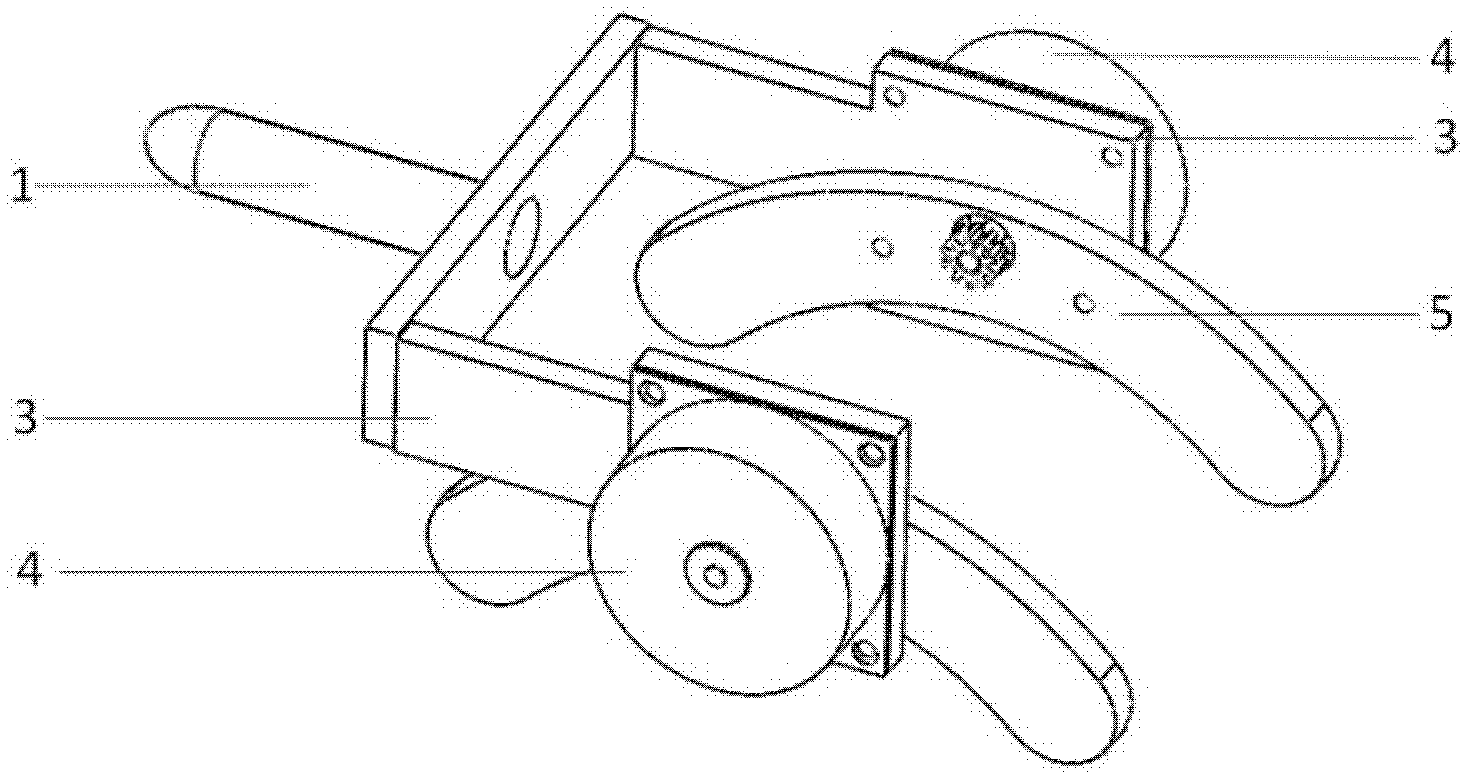
[0028] Such as figure 1 As shown, the three-dimensional ultrasonic imaging scanning device of this embodiment includes a handle 1, a probe 2, a side support plate 3, a stepping motor 4, an arc-shaped navigation track 5, an arc-shaped gear track 6, a driving gear 9, a probe clamp 7 and a fixed Connection plate 8. The arc navigation track 5, the arc gear track 6 and the driving gear constitute a swing mechanism.

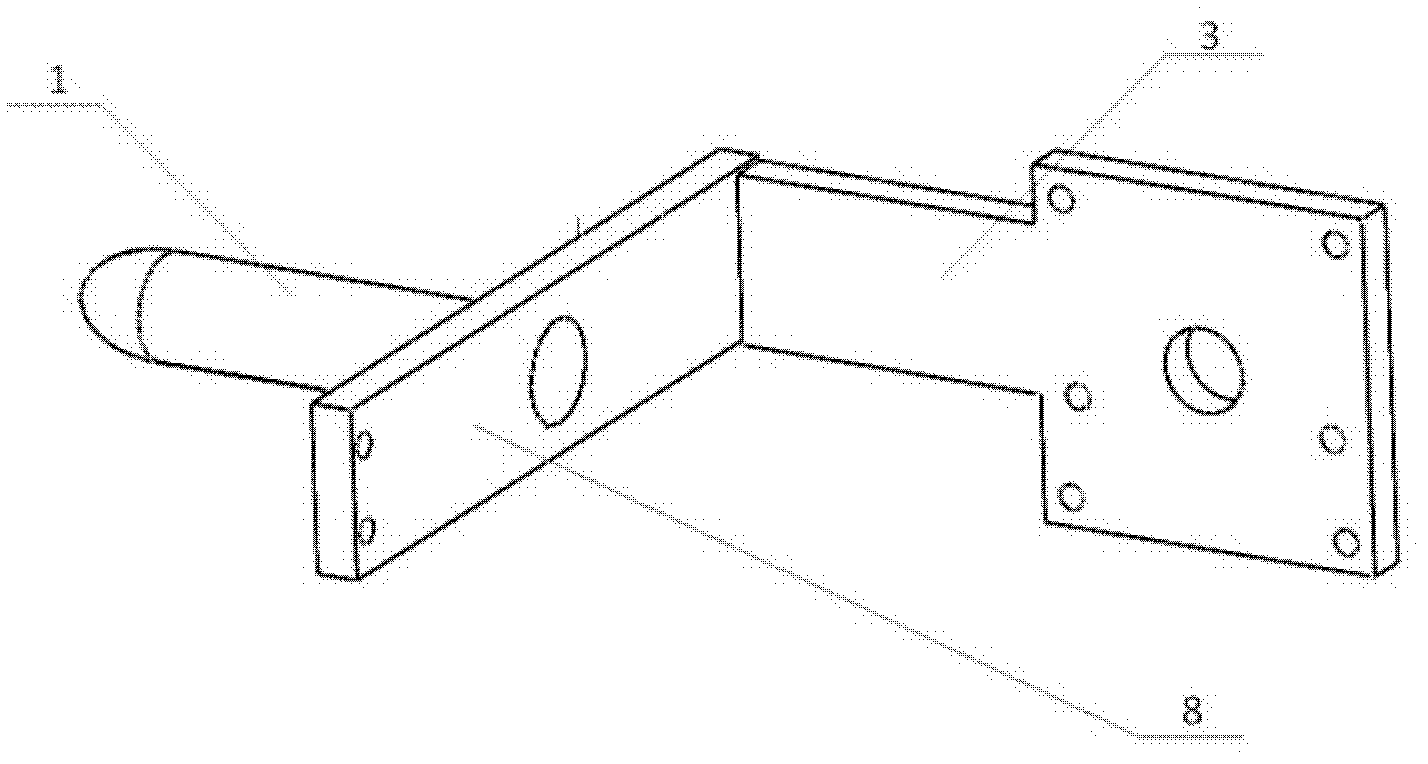
[0029] The bracket 3 includes two opposing and parallel side support plates, and the ends of one end of the parallel side support plates are connected by a fixed connecting plate 8 to form a whole, so that the bracket 3 as a whole has a "U"-shaped structu...


PUM


Abstract

The invention discloses a three-dimensional ultrasound image scanning device based on rear-end scanning, comprising a probe bracket, a probe clip (7), two stepper motors (4) and a swing mechanism. The probe bracket comprises two side support plates oppositely arranged in parallel; ends of the two side support plates in a same direction are connected through a fixed connecting plate into a whole; the probe clip (7) is arranged in the space between the two side support plates in a swinging manner and is provided with probes (2); the two stepper motors (4) are respectively arranged on the outer sides of the two side support plates for driving the probe clip (7) to swing in a bracket (3); and the swing mechanism is driven by the stepper motors (4) and drives the probe clip (7) to perform a sector scanning movement by adopting an ultrasonic wave emitting surface on the top of the probe (2) as a vertex so as to implement the collection of two-dimensional ultrasound data. The device is simple in structure, low in cost, reasonable in structure and good in imaging effect.

Description

technical field [0001] The invention belongs to the technical field of three-dimensional ultrasonic imaging, and in particular relates to a data acquisition device for three-dimensional ultrasonic images. Background technique [0002] The traditional B-mode ultrasound imaging system provides a two-dimensional image of a section of the human body. Doctors must synthesize multiple two-dimensional images in the brain based on their own experience to understand its three-dimensional anatomical structure. Compared with traditional two-dimensional ultrasound imaging, three-dimensional ultrasound imaging has obvious advantages. [0003] The basis of the current domestic three-dimensional ultrasound imaging is: based on the scanning data of the two-dimensional ultrasound probe and then obtained through the reconstruction algorithm. Sector scanning and scanning are the most frequently used scanning methods, but traditional sector scanning and scanning have inevitable defects: the an...

Claims


Application Information

Patent Timeline
IPC IPC(8): A61B8/00
Inventor 尉迟明丁明跃齐建
Owner 维视医学影像有限公司
Learn More