Multi-modality contrast and brightfield context rendering for enhanced pathology determination and multi-analyte detection in tissue

A technique for combining images, darkfield images, applied in the field of multimodal contrast and brightfield background reproduction for enhanced pathology assays and multianalyte detection of tissues, which can solve the problems of increased cost and time

Inactive Publication Date: 2012-09-12
VENTANA MEDICAL SYST INC
View PDF15 Cites 23 Cited by
  • Summary
  • Abstract
  • Description
  • Claims
  • Application Information

AI Technical Summary

Problems solved by technology

Comparing consecutive slices not only increases the expense and time necessary for the evaluation, it may be difficult or impossible to correlate features found in one slice with features found in another

Method used


Image

  • Multi-modality contrast and brightfield context rendering for enhanced pathology determination and multi-analyte detection in tissue
  • Multi-modality contrast and brightfield context rendering for enhanced pathology determination and multi-analyte detection in tissue
  • Multi-modality contrast and brightfield context rendering for enhanced pathology determination and multi-analyte detection in tissue

Examples


example

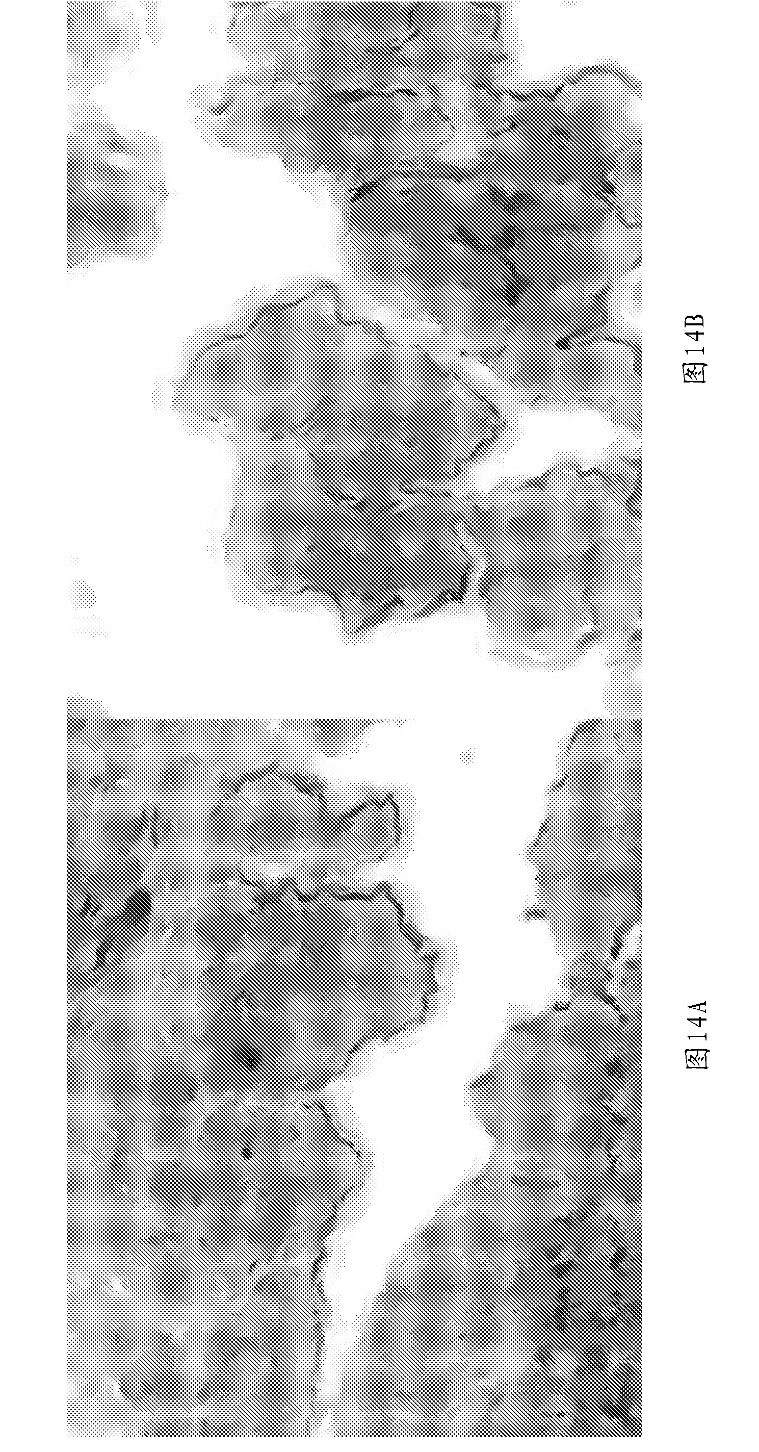
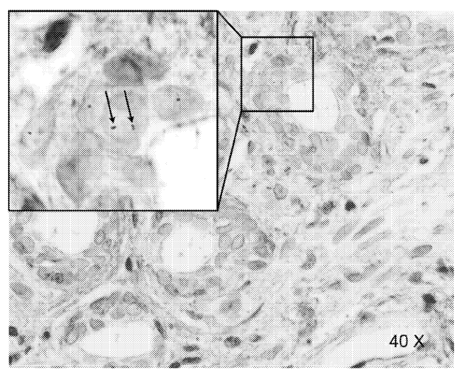
[0090] In some additional examples, images of formalin-fixed, paraffin-embedded histological tissue sections prepared according to the Ventana Medical System (Tucson, Arizona) protocol were obtained. In the examples of Figures 4, 5, 6, 10, 12, 13, 20, and 21, tissue sections were reconstructed from prostatectomy and processed with semiconductor nanocrystal quantum dots (QDots) for fluorescence in situ hybridization (FISH) , and counterstained with the fluorescent dye 4',6-diamidino-2-phenylindole (DAPI). QDot detection and DAPI fluorescence can be generated with a UV excitation beam in the wavelength range of 370 + / - 20 nm. Such an excitation beam is well suited for simultaneous multiple excitation of UV absorbing nuclear counterstains such as DAPI as well as multiple QDot probes. The refractive index contrast in the example contrast scheme is bright versus dark field and does not rely on light-absorbing staining, and thus allows simultaneous observation and recording of fluo...


PUM

PropertyMeasurementUnit
wavelengthaaaaaaaaaa

Abstract

Multiple modality contrast can be used to produce images that can be combined and rendered to produce images similar to those produced with wavelength absorbing stains viewed under transmitted white light illumination. Images obtained with other complementary contrast modalities can be presented using engineered color schemes based on classical contrast methods used to reveal the same anatomical structures and histochemistry, thereby providing relevance to medical training and experience. Dark-field contrast images derived from refractive index and fluorescent DAPI counterstain images are combined to produce images similar to those obtained with conventional H&E staining for pathology interpretation.; Such multi-modal image data can be streamed for live navigation of histological samples, and can be combined with molecular localizations of genetic DNA probes (FISH), sites of mRNA expression (mRNA-ISH), and immunohistochemical (IHC) probes localized on the same tissue sections, used to evaluate and map tissue sections prepared for imaging mass spectrometry.

Description

[0001] Cross References to Related Applications [0002] This application claims the benefit of U.S. Provisional Application No. 61,250,809, filed October 12, 2009, and U.S. Provisional Application No. 61 / 278,936, filed October 13, 2009, which are hereby incorporated by reference in their entirety. technical field [0003] The present disclosure relates to methods of providing comparison of tissue sections for pathological determination. Background technique [0004] Microscopic clinical examination of routine histologically stained tissue sections can be used to assess morphological patterns and tissue architecture of diagnostic significance. A skilled physician can observe such histologically stained tissue sections for diagnosis as well as for planning and evaluating treatment. The contrast of structures provided by such images using typical stains is familiar and allows the physician to devote herself to interpreting the features and abnormalities of the anatomical and ...

Claims


Application Information

Patent Timeline
Patent Type & Authority Applications(China)
IPC IPC(8): G01N33/483
CPCG01N21/6456
Inventor K.加沙G.佩斯塔诺M.奥特A.D.纳吉R.B.纳格勒P.米勒J.弗勒利希W.戴
Owner VENTANA MEDICAL SYST INC
Why Eureka
Try Eureka