Ultrasound diagnostic apparatus

A diagnostic device and ultrasonic technology, applied in the directions of sonic diagnosis, infrasonic diagnosis, ultrasonic/sonic/infrasonic diagnosis, etc., can solve problems such as the deterioration of ultrasonic image image quality

Inactive Publication Date: 2012-09-19
FUJIFILM CORP
View PDF9 Cites 8 Cited by
  • Summary
  • Abstract
  • Description
  • Claims
  • Application Information

AI Technical Summary

Problems solved by technology

[0011] In ultrasonic diagnostic equipment, the so-called near field is more likely to deteriorate the image quality of ultrasonic images due to sound speed disturbance and multiple refraction

Method used


Image

  • Ultrasound diagnostic apparatus
  • Ultrasound diagnostic apparatus
  • Ultrasound diagnostic apparatus

Examples


Embodiment Construction

[0055] Hereinafter, the ultrasonic diagnostic apparatus of the present invention will be described in detail with reference to preferred embodiments shown in the accompanying drawings.

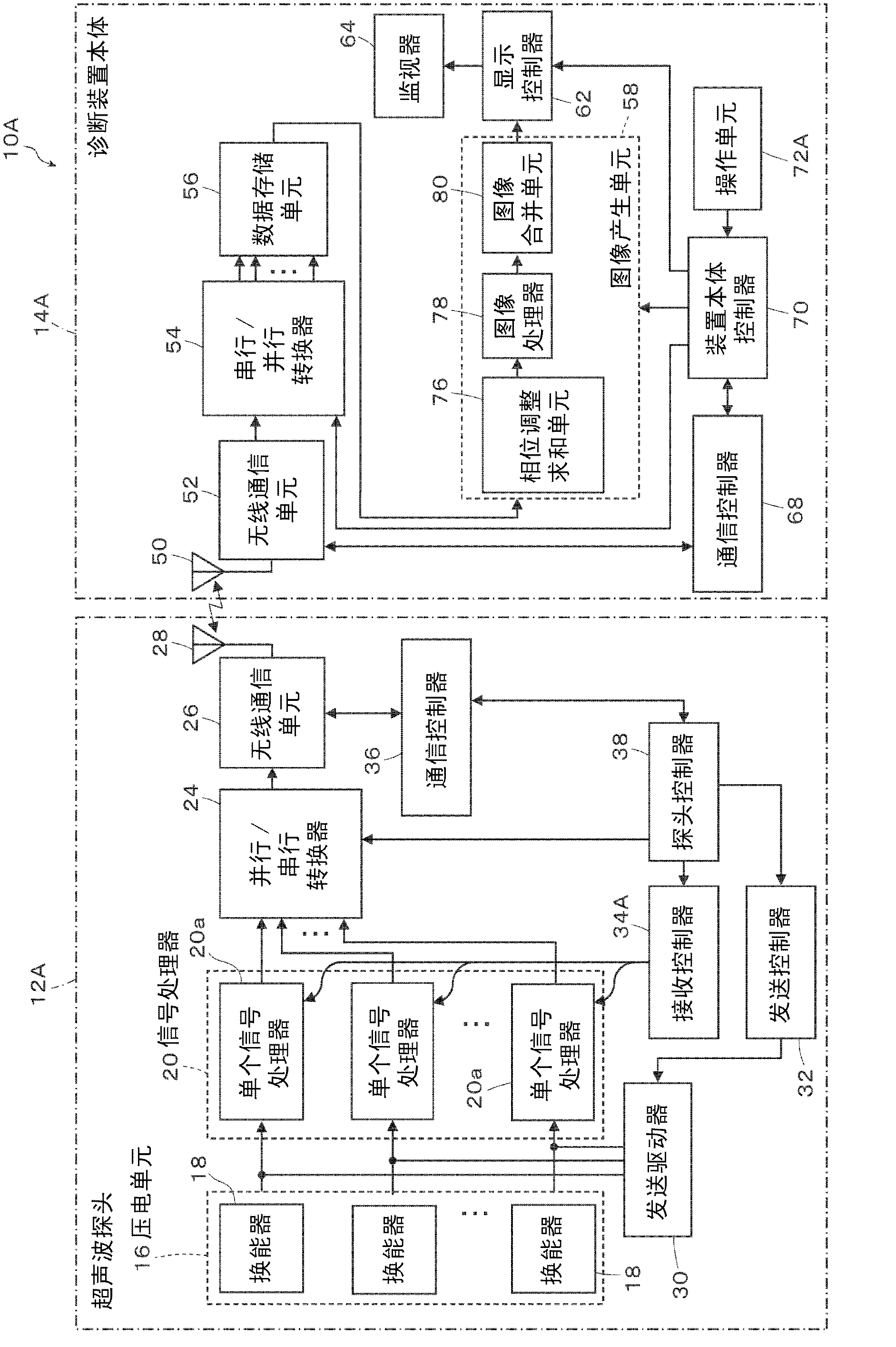
[0056] figure 1 is a schematic block diagram of an embodiment of the ultrasonic diagnostic apparatus according to the first aspect of the present invention.

[0057] figure 1 The illustrated ultrasonic diagnostic apparatus 10A includes an ultrasonic probe 12A and a diagnostic apparatus body 14A. The ultrasound probe 12A is connected to the diagnostic apparatus main body 14A in a wireless communication manner.

[0058] The ultrasonic probe 12A (hereinafter referred to as "probe 12A") transmits ultrasonic waves to a subject, receives ultrasonic echoes generated by the ultrasonic waves reflected on the subject, and outputs reception signals of ultrasonic images based on the received ultrasonic echoes.

[0059] In the implementation of the present invention, various known ultrasonic probes ca...


PUM


Abstract

An ultrasound diagnostic apparatus is provided which includes an ultrasound probe performing ultrasound transmission and reception in different directions and a diagnostic apparatus body combining images different in the direction of transmission and reception to produce an ultrasound image. At least one of ultrasound images to be combined is changed depending on the set region of interest (ROI) to an image of the depth corresponding to the ROI. When an acoustic coupler is attached, the depth of at least one of ultrasound images to be combined is increased. The ultrasound diagnostic apparatus is capable of improving the image quality of the ROI and obtaining efficient high-definition ultrasound images reaching a predetermined depth when the acoustic coupler is attached.

Description

technical field [0001] The present invention relates to ultrasonic diagnostic devices. More specifically, the present invention relates to an ultrasonic diagnostic apparatus capable of improving the image quality of a region of interest and obtaining a high-definition ultrasonic image that efficiently shows the vicinity of a subject's skin surface. Background technique [0002] Ultrasonic diagnostic devices using ultrasonic images have been put into practical use in the medical field. [0003] In general, an ultrasonic diagnostic device includes an ultrasonic probe (hereinafter referred to as "probe") and a diagnostic device body. In an ultrasonic diagnostic apparatus, a probe transmits ultrasonic waves to a subject and receives ultrasonic echoes from the subject. The diagnostic device body electrically processes reception signals received by and output from the probe to generate ultrasonic images. [0004] The probe performs transmission and reception of ultrasonic waves...

Claims


Application Information

Patent Timeline
Patent Type & Authority Applications(China)
IPC IPC(8): A61B8/00
CPCA61B8/4281A61B8/56A61B8/469A61B8/5253G01S7/52085A61B8/4472G01S15/8995A61B8/546
Inventor 田边刚大岛雄二
Owner FUJIFILM CORP
Why Eureka
Try Eureka