Real-time amplification method for ultrasonic image

An ultrasound image and ultrasound technology, applied in image data processing, graphic image conversion, instruments, etc., can solve the problems of small dynamic range of data, unable to present image details, small dynamic range of images, etc., to achieve the effect of improving quality

Active Publication Date: 2012-09-19
VINNO TECH (SUZHOU) CO LTD
View PDF4 Cites 11 Cited by
  • Summary
  • Abstract
  • Description
  • Claims
  • Application Information

AI Technical Summary

Problems solved by technology

[0008] (1) When the main image and the enlarged image are displayed at the same time, they share the same original data source. Zoom in to the specified multiple, this method is simple to implement, but reduces the quality of the enlarged image, which is not conducive to detailed diagnosis;
[0009] (2) Using real-time sound amplification technology to amplify the ultrasonic image, the current method of increasing sampling lines and sampling points cannot effectively improve

Method used


Image

  • Real-time amplification method for ultrasonic image
  • Real-time amplification method for ultrasonic image
  • Real-time amplification method for ultrasonic image

Examples


Embodiment Construction

[0026] The structure of the present invention will be further described below in conjunction with the accompanying drawings.

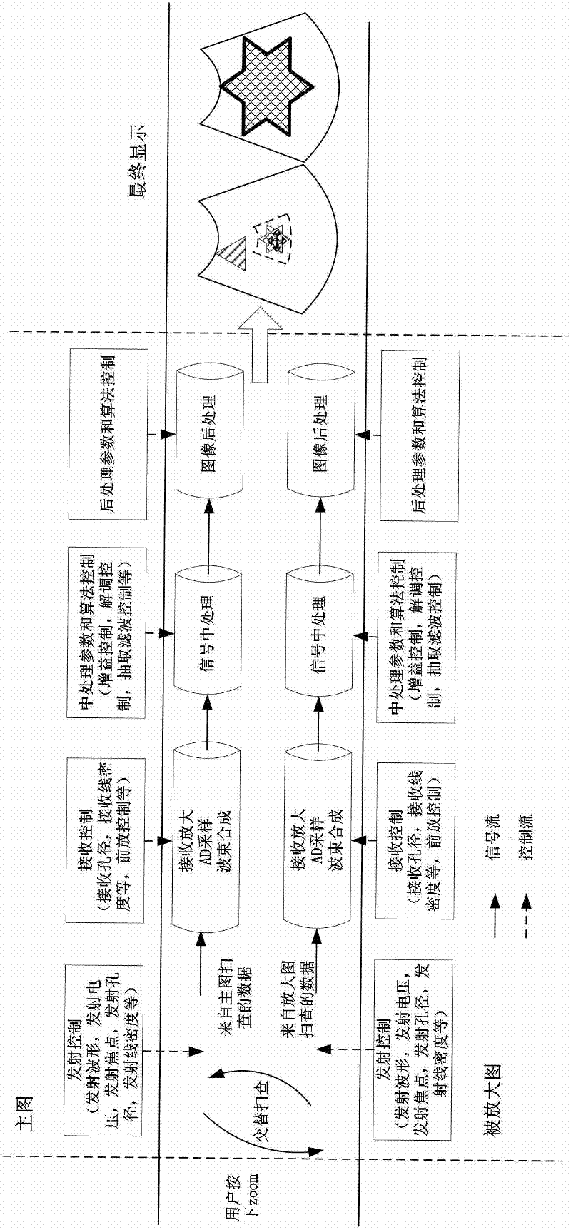
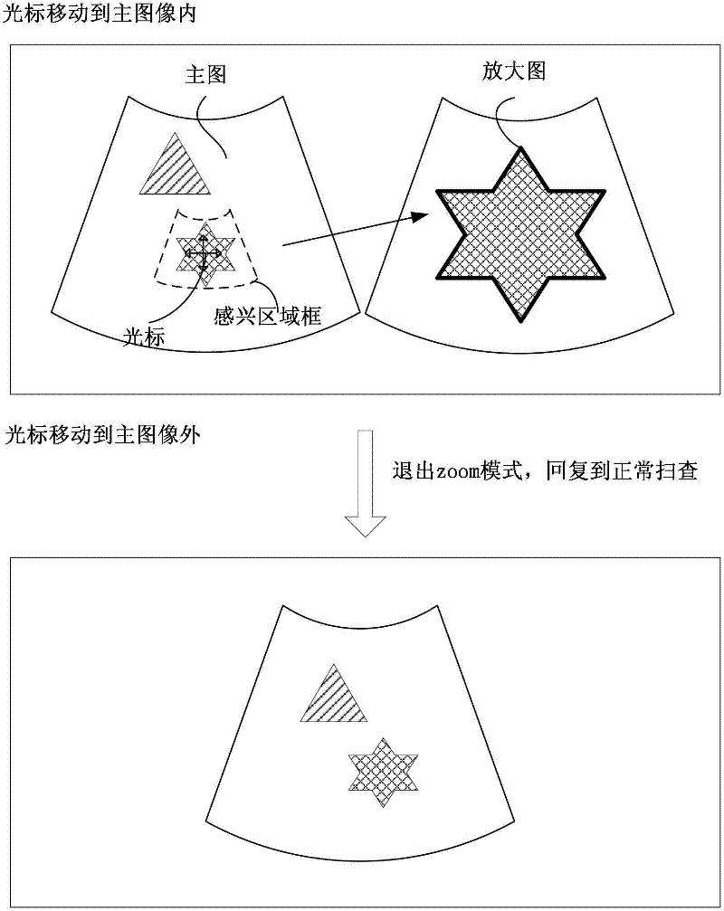
[0027] A method for real-time magnification of an ultrasound image, comprising a movable region-of-interest frame appearing on a main image to obtain an enlarged image corresponding to the region-of-interest frame, the ultrasonic scanning of the main image and the ultrasonic scanning of the enlarged image are independent of each other Yes, when entering the zoom mode, the scanning timing of the main image and the scanning timing of the enlarged area (region of interest) can be completely independently controlled. Use S main Characterize the main Figure 1 Frame scan, use S zoom It represents the scanning of one frame in the enlarged area. In the simultaneous mode, if the doctor wants to see the real-time main image and the enlarged image at the same time, the alternate scanning method is used.

[0028] S main ,S zoo...


PUM


Abstract

The invention discloses a real-time amplification method for an ultrasonic image. A main image and an amplified image are subjected to ultrasonic scanning control and scanning imaging chain processing which are independent from each other, so that not only can the sampling linear density and the sampling rate of the amplified image be improved, but also parameter and signal treatment capable of improving resolution ratios and contrast ratios of the amplified image can be applied to the amplified image on the whole imaging chain, so that compared with an existing method of utilizing an image post-treatment interpolation or adding a sampling line and a sampling point at the front end to improve the quality of the amplified image, the real-time amplification method can more completely and more effectively improve the quality of the amplified image to the greater extent. The real-time amplification method for the ultrasonic image further adopts a compression method depending on data in an area-of-interest to present information in the area-of-interest in an expanded dynamic range, so that more details of the amplified image can be observed.

Description

[0001] technical field [0002] The invention relates to a real-time zoom-in method for medical ultrasonic images. Background technique [0003] Ultrasound image magnification plays an important role in the doctor's diagnosis of details. At present, the zoom-in function of most ultrasound imaging systems is mainly based on image post-processing, that is, the zoom-in is performed through image processing according to the region of interest selected by the user. In terms of workflow, the user has to press "zoom" first, and then a region of interest frame appears, and the user can change the position of the region of interest frame by moving the magic ball, so that the enlarged image is displayed on the main interface. The enlarged image may be a pop-up, or the enlarged image may appear in the corresponding position of the main image and so on. However, since the magnified image and the main image share the same original data source, the magnified image is only a local image ...

Claims


Application Information

Patent Timeline
IPC IPC(8): G06T3/40
Inventor 吴方刚
Owner VINNO TECH (SUZHOU) CO LTD
Why Eureka
Try Eureka