Improved region growing method applied to coronary artery angiography image segmentation

A technique for angiographic images and coronary arteries, which is applied in the field of image processing, can solve problems such as undergrowth, increased calculation, and difficulty in segmenting images with uneven brightness, and achieve the effect of improving accuracy and speed

Inactive Publication Date: 2012-10-17
常熟市支塘镇新盛技术咨询服务有限公司
View PDF1 Cites 21 Cited by
  • Summary
  • Abstract
  • Description
  • Claims
  • Application Information

AI Technical Summary

Problems solved by technology

[0006] 2. The growth criterion of traditional region growth is judged according to the similarity of adjacent pixels or the average pixel gray value. This criterion is difficult to segment images with uneven brightness
like figure 1 , the background at A is larger than the gray level of blood vessels at B, using similarity or global average gray level, it is easy to produce overgrowth or undergrowth
There are also some methods that use the statistical information of pixels as the growth criterion, but the amount of calculation of those methods is greatly increased.

Method used


Image

  • Improved region growing method applied to coronary artery angiography image segmentation
  • Improved region growing method applied to coronary artery angiography image segmentation
  • Improved region growing method applied to coronary artery angiography image segmentation

Examples


Embodiment Construction
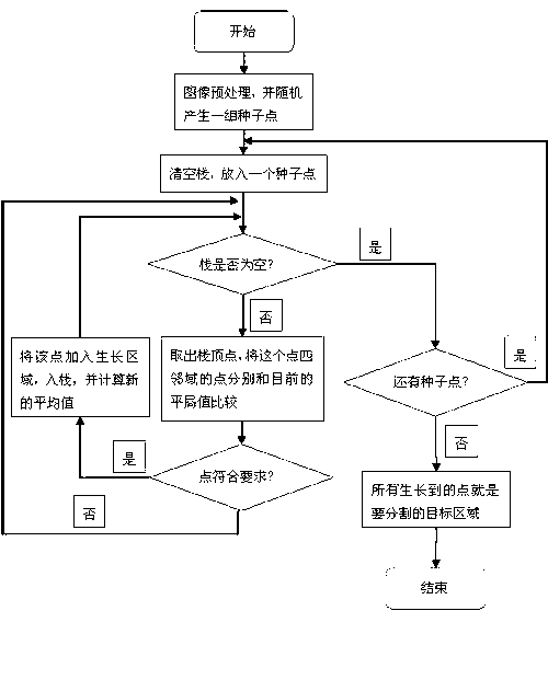
[0020] The basic idea of ​​region growth is to group pixels with similar properties to form regions. Specifically, first find a seed pixel for each region to be segmented as the starting point of growth, and then merge the pixels that have the same or similar properties as the seed pixel in the area around the seed pixel into the area where the seed pixel is located. Treat these new pixels as new seed pixels and continue the above process until no more pixels that meet the conditions can be included. Combine below figure 1 The specific steps of image segmentation in the present invention will be described in detail.

[0021] figure 2 It is a pre-processed coronary angiogram, and now its width is M and height is N. Use G(i, j) to represent the gray value of the pixel with coordinates (i, j), and give each point a mark F i, j , F i, j = 0 means that the point (i, j) does not belong to the growing area, F i, j = 1 means the point is already grown.

[0022] Step 1: Automatic...


PUM


Abstract

The invention relates to an improved region growing method which is applied to vessel segmentation and extraction in a coronary artery angiography image. The improved region growing method comprises the following steps of: preprocessing the image to obtain an original image capable of directly performing region growth; making a regulation and randomly generating a group of seed points; setting a stack data structure, enabling a newly grown pixel point to enter a stack, and taking out the point previously entering the stack to serve as a current point to be subjected to growth when the current point completes the growth; sequentially performing growth on each seed point, wherein a seed point gray value serves as an average value at a growing initial stage, and calculating a new average gray value when a new pixel point is grown every time along with the growth of the seed points; and completing the growth when no pixel point meeting growth standards exists and no seed point exists. The improved region growing method has the advantages that the seed points are automatically generated, no manual intervention is needed, the local average values around each pixel point serve as growth parameters in a growing process, the coronary artery angiography image with uneven brightness can be segmented, and the efficiency and the accuracy of the image segmentation are improved.

Description

1. Technical Field [0001] The present invention relates to image processing, in particular to an improved region growing method used for segmentation and extraction of blood vessels in coronary angiography images. 2. Background technology [0002] Medical image processing is an important branch in the field of image processing, and the segmentation and extraction of blood vessels is one of the key and difficult points of medical image processing. Coronary angiography is to insert a catheter after percutaneously piercing an artery, and inject a contrast agent, using X-rays to absorb the contrast agent and other human tissues to record the image of the blood vessel. Therefore, the image brightness is very uneven. Such as figure 1 Shown is a smooth filtered coronary angiogram, the brightness of the image is not uniform. [0003] The region growing algorithm was first proposed by Zucker. As a serial region algorithm, it starts from one or a set of starting seed points, and continuous...

Claims


Application Information

Patent Timeline
Patent Type & Authority Applications(China)
IPC IPC(8): G06T7/00
Inventor 刘向荣於猛黄晓阳
Owner 常熟市支塘镇新盛技术咨询服务有限公司
Why Eureka
Try Eureka