Ultrasound diagnostic apparatus and ultrasound diagnostic apparatus control method

A diagnosis device and control method technology, applied in the direction of ultrasonic/sonic wave/infrasonic equipment control, sound wave diagnosis, infrasonic wave diagnosis, etc., can solve problems such as inability to measure correctly, and achieve the effect of good operation feeling

Active Publication Date: 2012-12-19
KONICA MINOLTA INC
View PDF8 Cites 9 Cited by
  • Summary
  • Abstract
  • Description
  • Claims
  • Application Information

AI Technical Summary

Problems solved by technology

) in the vicinity of the center of the ultrasonic wave, if the lumen-intima boundary 204 and media-adventi

Method used


Image

  • Ultrasound diagnostic apparatus and ultrasound diagnostic apparatus control method
  • Ultrasound diagnostic apparatus and ultrasound diagnostic apparatus control method
  • Ultrasound diagnostic apparatus and ultrasound diagnostic apparatus control method

Examples


Example Embodiment

[0040] Hereinafter, embodiments of the ultrasonic diagnostic apparatus and the control method of the ultrasonic diagnostic apparatus of the present invention will be described in detail with reference to the drawings.
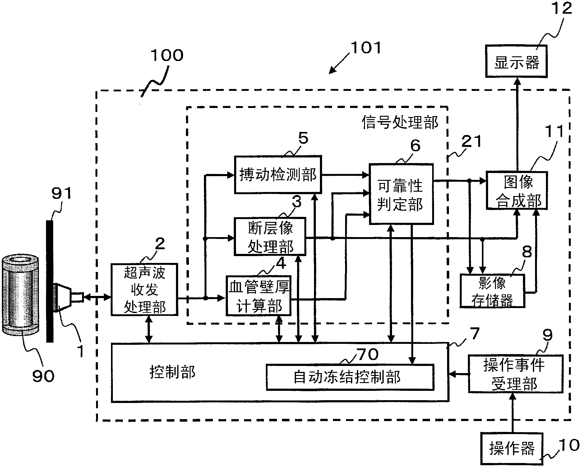
[0041] In this embodiment, the ultrasonic diagnostic apparatus performs IMT measurement of the subject. figure 1 It is a block diagram showing the ultrasonic diagnostic apparatus of this embodiment.

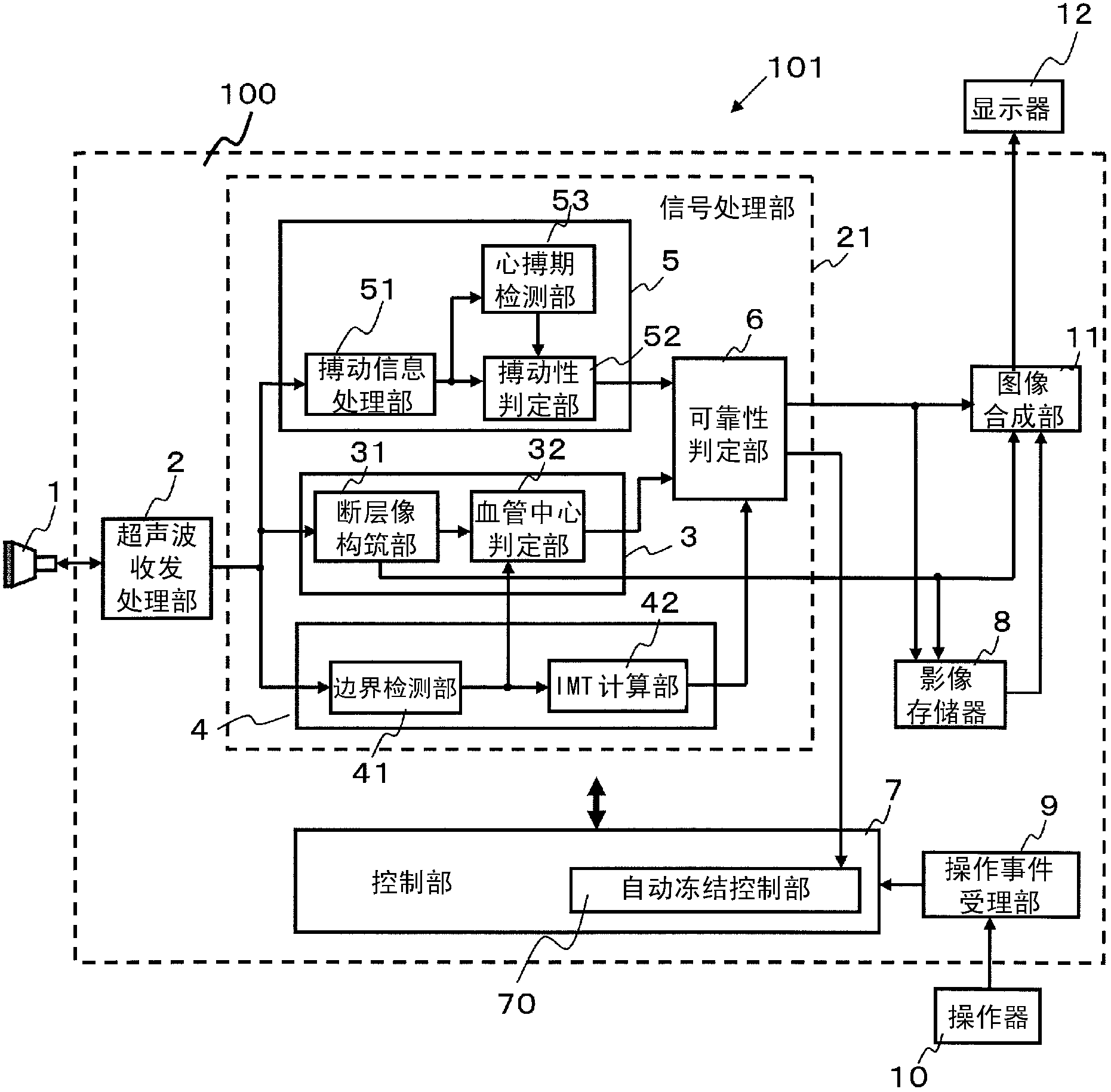
[0042] The ultrasonic diagnostic apparatus 101 includes a controller 100. The controller 100 includes an ultrasonic transmission and reception processing unit 2, a signal processing unit 21, a control unit 7, an image memory 8, an operation event receiving unit 9 and an image synthesis unit 11. In this embodiment, the ultrasonic diagnostic apparatus 101 may not be equipped with the probe 1, but a general-purpose probe 1 may be connected. However, the ultrasonic diagnostic apparatus 101 may be equipped with the probe 1.

[0043] The ultrasonic transmission / reception pr...


PUM


Abstract

This ultrasound diagnostic apparatus, to which a probe having a transducer can be connected, is provided with a controller (100) that: performs a transmission process that sends ultrasonic waves into a subject by driving a probe (1), and a reception process that generates a received signal based on reflected ultrasonic waves received by said probe from the subject; and when a prescribed condition based on the received signal is satisfied, performs an automatic freeze process, wherein at least one of the signal transmission process or reception process is stopped. The controller (100) inhibits the automatic freeze process for a prescribed period after receiving a prescribed operation event.

Description

technical field [0001] The present invention relates to an ultrasonic diagnostic device and a control method for the ultrasonic diagnostic device. Background technique [0002] Ultrasonic diagnostic apparatuses are capable of acquiring tomographic images in the subject's body, and are widely used for shape observation of tissues in the body, and the like. In addition, in the diagnosis of arteriosclerosis, it is also used to measure the intima-media thickness (Initima-Media Thickness, hereinafter abbreviated as IMT) of the carotid artery, which is an important index for understanding early stage atherosclerosis. IMT is the thickness of the intima-media complex of the carotid artery wall, such as Figure 11 (a) and (b) refer to the thickness of the layer (intima-media 203 ) seen between the lumen 201 and the adventitia 202 of the blood vessel. The ultrasonic diagnostic apparatus detects the boundary between the lumen 201 and the intima (hereinafter referred to as the lumen-in...

Claims


Application Information

Patent Timeline
IPC IPC(8): A61B8/06A61B8/00
CPCA61B8/461A61B8/145A61B8/4488A61B8/5223A61B8/483A61B8/5207A61B8/4444A61B8/0891A61B8/54G16H50/30
Inventor 川端章裕伊藤嘉彦铃木隆夫
Owner KONICA MINOLTA INC
Why Eureka
Try Eureka