Load small interfering RNA nanoscale lipid microbubble ultrasonic contrast agent and preparation method
A technology of ultrasonic contrast agent and lipid microbubble, which is applied in the field of ultrasonic molecular imaging and biomedical engineering, can solve the problems of large diameter, ineffective combination, and inability to load gene fragments, etc., to achieve integration and good enhancement of ultrasonic imaging effect of ability
- Summary
- Abstract
- Description
- Claims
- Application Information
AI Technical Summary
Problems solved by technology
Method used
Image
Examples
Embodiment 1
[0035] S1. Preparation of nano-scale lipid microbubble ultrasound contrast agent (hereinafter referred to as nano-microbubble) with negatively charged surface:
[0036] Nano-microbubbles were prepared using phospholipids as raw materials by thin film-hydration method. The phospholipid components dipalmitoylphosphatidylcholine (DPPC), distearoylphosphatidylethanolamine (DSPE), dipalmitoylphosphatidic acid (DPPA) were dissolved in chloroform in parts by weight 18:1:1 and placed in diameter 9 cm petri dish, in the fume hood to wait for the chloroform to volatilize naturally to form a phospholipid film. After adding 4 ml of double-distilled water and hydrating at 37 °C for 1 hour, the lipid solution was transferred to a 50 ml centrifuge tube, sonicated for 5 minutes with an ultrasonic breaker, and at the same time, octafluoropropane gas was introduced to prepare lipid microbubbles. The microbubble liquid was left to stand for 10 minutes, and then centrifuged at 1000 rpm for 5 min...
Embodiment 2
[0044] Example 2 Diameter and Surface Potential of Intermediate and Final Products:
[0045] The diameters and surface potentials of the intermediate products (nanobubbles, siRNA micelles) and final products (siRNA nanobubbles) obtained in Example 1 were detected by dynamic light scattering. The results (result data expressed as mean ± standard error) show that the diameters of nanobubbles, siRNA micelles, and siRNA nanobubbles are 436.8±5.7 nm, 66.8±2.8 nm, and 476.0±6.1 nm, respectively; and the surface potential of the three -18.4±0.2 mV; 23.4±1.1 mV, 15.3±2.7 mV. The diameter and surface potential verified the polymerization process of preparing siRNA nano-microbubbles.
Embodiment 3
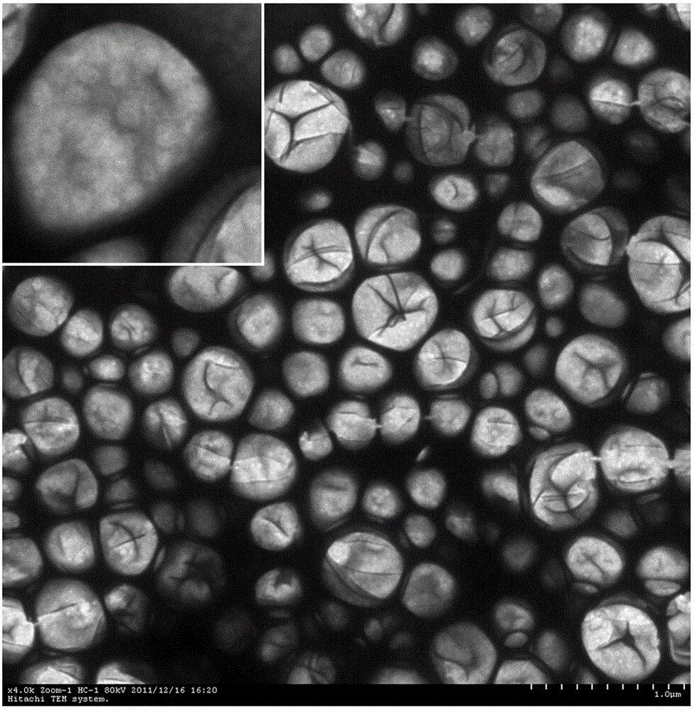
[0046] Example 3 Morphological structure detection of siRNA nano-microbubbles:
[0047] In order to further confirm the morphology and structure of the resulting siRNA nano-microbubbles prepared in Example 1, a transmission electron microscope is used to detect, and the test results are shown in figure 1 . Through the test, it can be seen that the siRNA nano-microbubbles are round or quasi-circular, and the diameter distribution is about 200-500 nm. The diameter distribution is uniform and there is no obvious aggregation. The microbubble surface dents caused by vacuuming during the preparation of the transmission electron microscope can be seen on the surface . Visible at high magnification ( figure 1 Upper left corner), the contrast agent is approximately round, the surface is not smooth, and a large number of small siRNA micelles with a diameter of about 50-70 nm can be seen on the surface of the microbubbles to form an overall structure.
PUM
| Property | Measurement | Unit |
|---|---|---|
| diameter | aaaaa | aaaaa |
| diameter | aaaaa | aaaaa |
| diameter | aaaaa | aaaaa |
Abstract
Description
Claims
Application Information
 - R&D
- Intellectual Property
- Life Sciences
- Materials
- Tech Scout
- Unparalleled Data Quality
- Higher Quality Content
- 60% Fewer Hallucinations
Browse by: Latest US Patents, China's latest patents, Technical Efficacy Thesaurus, Application Domain, Technology Topic, Popular Technical Reports.
© 2025 PatSnap. All rights reserved.Legal|Privacy policy|Modern Slavery Act Transparency Statement|Sitemap|About US| Contact US: help@patsnap.com


