Automatic identification of intracardiac devices and structures in an intracardiac echo catheter image

An image and catheter technology, applied in the field of medical imaging and physiological modeling, can solve problems such as distortion, image complexity, echo image cannot be represented, etc.

Active Publication Date: 2013-09-04
ST JUDE MEDICAL ATRIAL FIBRILLATION DIV
View PDF13 Cites 4 Cited by
  • Summary
  • Abstract
  • Description
  • Claims
  • Application Information

AI Technical Summary

Problems solved by technology

However, the navigation electric field is not guaranteed to be uniform or isotropic, so for these geometries, distortions are often
The positioning of ICE catheters and the images they generate is further complicated by the fact that echo images often do not represent the idealized echo plane, as it has been observed that echo images often exhibit rotational and translational deviations from the ideal

Method used


Image

  • Automatic identification of intracardiac devices and structures in an intracardiac echo catheter image
  • Automatic identification of intracardiac devices and structures in an intracardiac echo catheter image
  • Automatic identification of intracardiac devices and structures in an intracardiac echo catheter image

Examples


Embodiment Construction

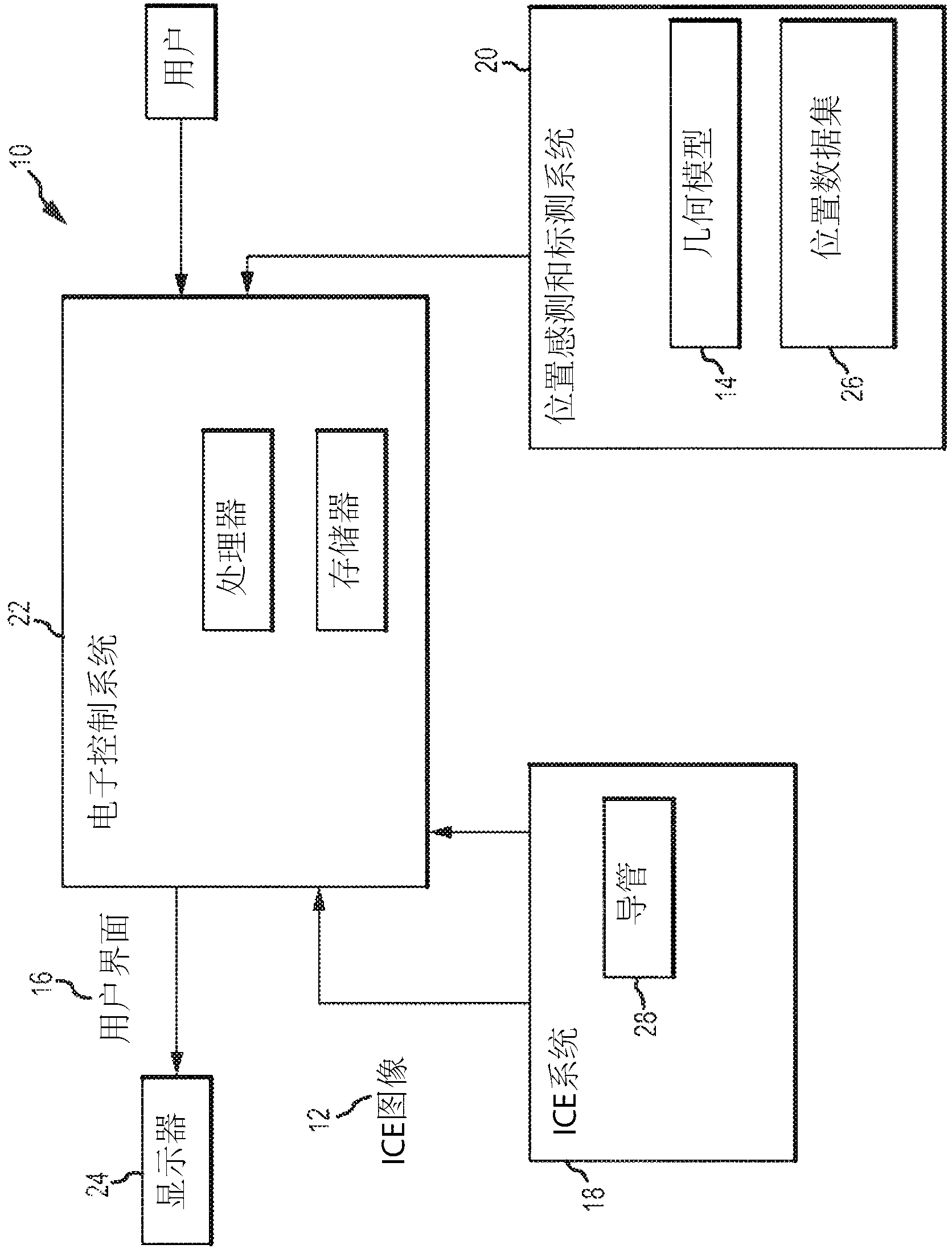
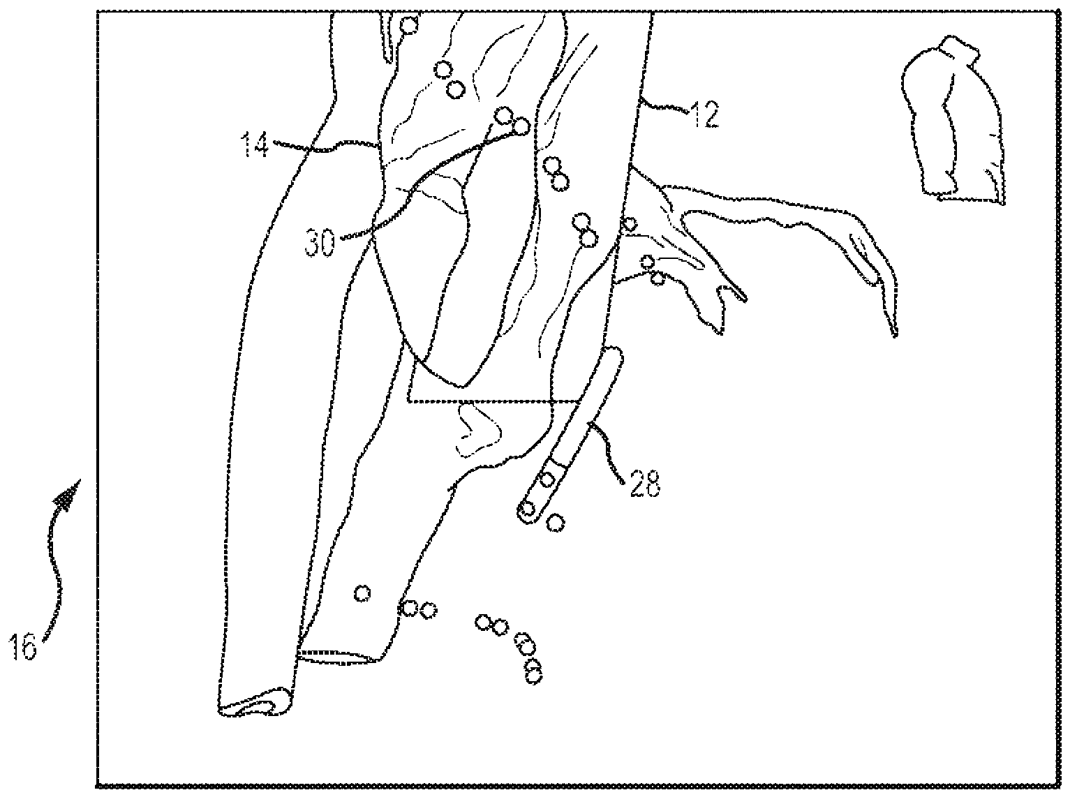
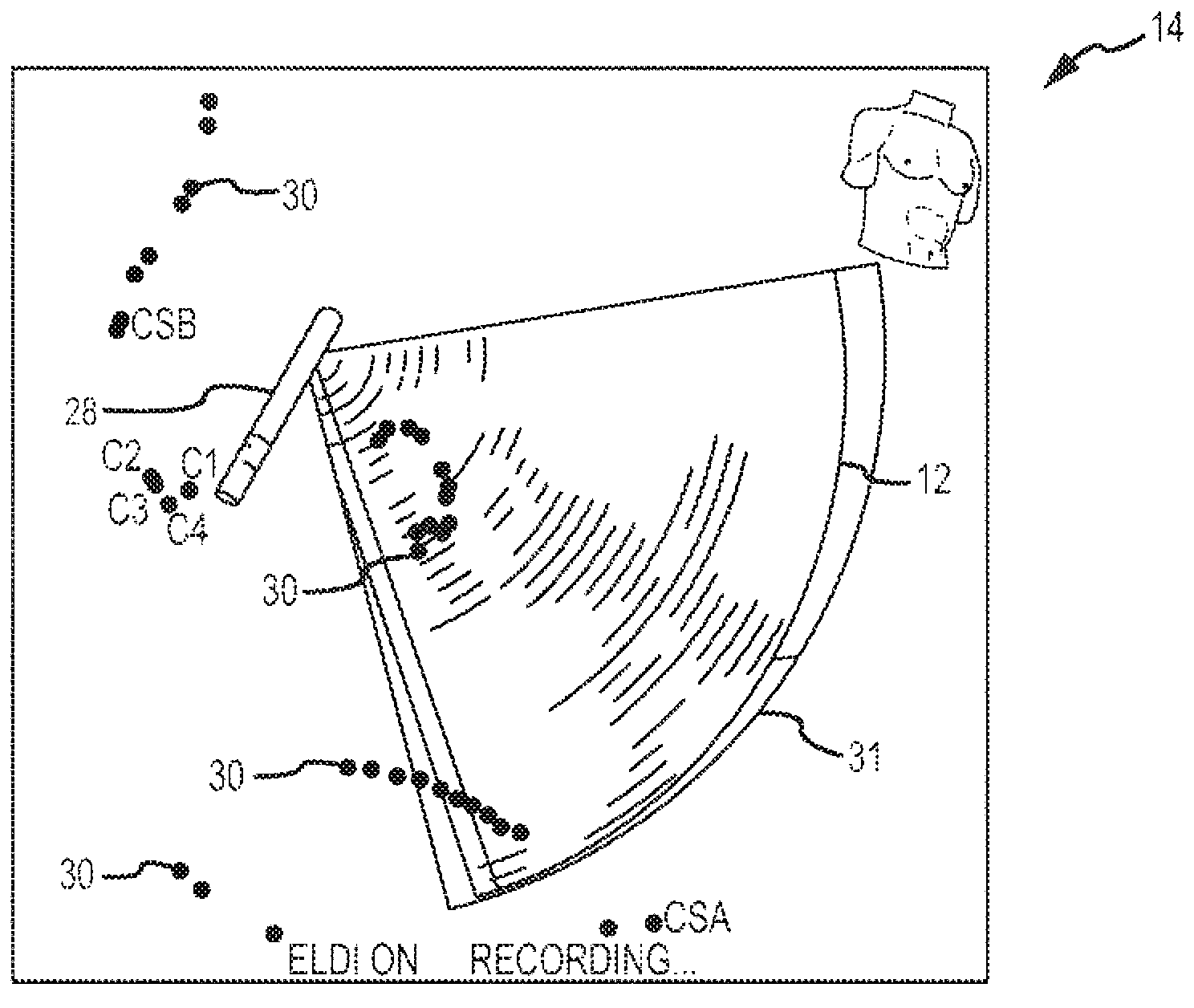
[0020] Referring now to the drawings, where the same reference numerals are used in different views to denote the same component, figure 1 An exemplary embodiment of the system 10 is illustrated. The system 10 is configured to display devices present in the geometric model 14 of the heart within an intracardiac echocardiographic image 12 (ICE image), and automatically segment the ICE image 12 to generate one or more 壳unit 36. The system 10 is further configured to generate a user interface 16 to display the ICE image 12 and geometric model 14 and to receive user input to guide the control and operation of the system 10.

[0021] The system 10 according to one embodiment of the present disclosure includes an intracardiac echo imaging system 18 (ICE system), a visualization, navigation, or mapping system 20 (“VNM” system), an electronic control system (ECS) 22, and a display 24. The ECS 22 may be configured to receive the ICE image 12 generated by the ICE system 18, and the ECS 22 ...


PUM


Abstract

An intracardiac imaging system 10 configured to display electrode visualization elements within an intracardiac echocardiography image 12 where the electrode visualization elements represent intracardiac electrodes in close proximity to the plane of the image 12. The system 10 further allows cross sections of tissue structures embodied in intracardiac echocardiography images 12 to be modeled within a visualization, navigation, or mapping system 20 when automatically segmented to generate shell elements 36 for modifying the modeled tissue structures.

Description

[0001] Cross references to related applications [0002] This application claims the priority of U.S. Patent Application 12 / 983,013 filed on December 31, 2010, which is incorporated herein by reference as if fully stated herein. Technical field [0003] The present invention relates to medical imaging and physiological modeling, and in particular, the present invention relates to the identification and tracking of equipment and structures in an imaging or modeling mode, and the simultaneous display of those information in a separate imaging or modeling mode. Background technique [0004] It is well known that the prevalence of different imaging modalities in the medical field provides clinicians with valuable information about patient physiology. However, all imaging modes have some type of error, which introduces uncertainty into the generated image. Further limiting the application of medical images is that when the image does not contain recognizable landmarks to provide a backg...

Claims


Application Information

Patent Timeline
Patent Type & Authority Applications(China)
IPC IPC(8): A61B8/12
CPCA61B8/0841A61B8/0883A61B8/12A61B8/4245A61B8/461A61B8/463A61B8/5246G06T19/00G06T2210/41
Inventor A·希尔D·C·德诺H·钟M·M·格拉斯
Owner ST JUDE MEDICAL ATRIAL FIBRILLATION DIV
Why Eureka
Try Eureka