Quick Monte Carlo imaging method for reconstructing optical parameter of tissue with heteroplasmon

A technology of optical parameters and imaging methods, which is applied in the field of biomedical engineering, can solve the problems of great differences, occupying a large storage space, and increasing the calculation time of the reconstruction process, so as to shorten the calculation time and save storage space.

Inactive Publication Date: 2013-10-23
TIANJIN UNIV
View PDF7 Cites 26 Cited by
  • Summary
  • Abstract
  • Description
  • Claims
  • Application Information

AI Technical Summary

Problems solved by technology

However, due to the need to run a complete MC simulation to obtain the path of the photon in the tissue to obtain k and s, the current pMC algorithm is limited to the reconstruction of the optical parameters of the homogeneous medium, or the position of the heterogeneous body is known. Reconst

Method used


Image

  • Quick Monte Carlo imaging method for reconstructing optical parameter of tissue with heteroplasmon
  • Quick Monte Carlo imaging method for reconstructing optical parameter of tissue with heteroplasmon
  • Quick Monte Carlo imaging method for reconstructing optical parameter of tissue with heteroplasmon

Examples


Embodiment Construction

[0035] The present invention will be described below in conjunction with accompanying drawing and embodiment

[0036] 1. Use the reciprocity method to improve the calculation efficiency of the number of collisions k and the distance traveled s

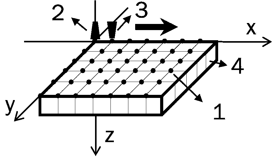
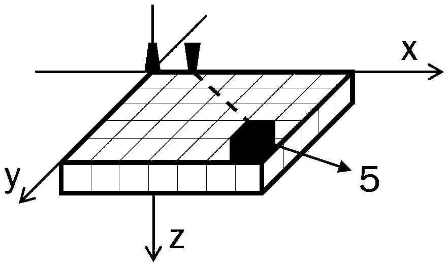
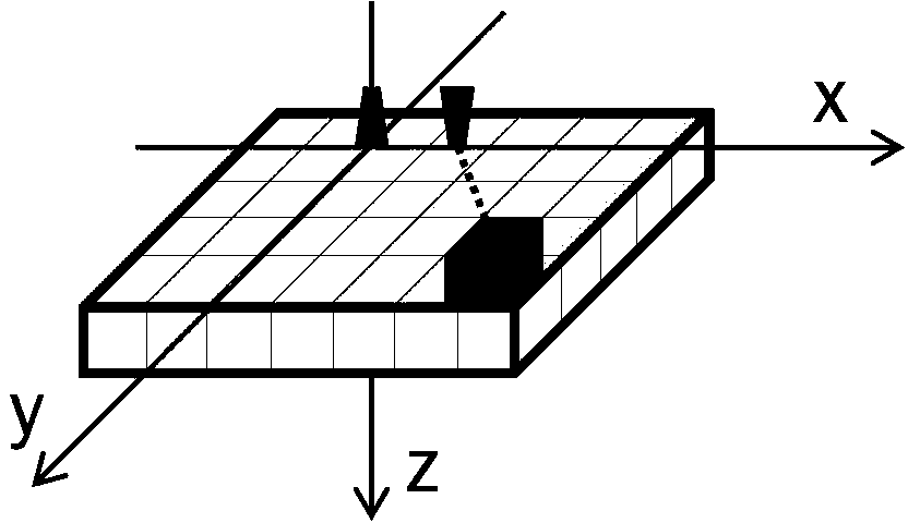
[0037] For biological tissues containing tumors, it can be approximately replaced by a flat plate model with heterogeneity with the same optical parameters (as shown in Figure (1)), and the positions of the light source and detector are set as shown in the figure. In order to obtain the distribution of optical parameters in the model, thereby obtaining the position of the heterogeneous body and its optical parameters, the flat plate model needs to be split axially (see Figure (1)) and divided into a series of homogeneous voxels. Select the intersection of the subdivision grid lines as the scanning point, make the light source scan along the scanning point line by line in the direction of the arrow shown in the figure, and keep the rela...


PUM


Abstract

The invention belongs to the technical field of biomedical engineering, and relates to a quick Monte Carlo imaging algorithm for reconstructing an optical parameter of a tissue with a heteroplasmon. The algorithm comprises the steps of confirming a detection zone, axially subdividing the detection zone, selecting intersection points of subdivision grid lines as scanning points, obtaining diffuse reflection light distribution in different scanning positions, performing MC (Monte Carlo) simulation when an illuminant is located at an original point and detectors are symmetrically distributed on the two sides of the illuminant, and obtaining mean collision times and mean walking lengths of photons in volume elements and photon weights received by all the detectors, Step III, restoring the collision times and the walking lengths in the volume elements when source detectors are positioned in all the scanning points, calculating and outputting weights of the photons, and determining a Jacobi matrix, Step IV, working out a position and the optical parameter of the heteroplasmon. The method has the benefits that the optical parameter of the tissue with the heteroplasmon can be reconstructed; the pMC (perturbation Monte Carlo) algorithm is optimized; a memory space is saved; and the calculation time is shortened.

Description

technical field [0001] The invention belongs to the technical field of biomedical engineering and relates to a diffuse light functional tomography algorithm suitable for mesoscopic and medium spatial resolution. Background technique [0002] Cancer can not only lead to changes in tissue structure and cell morphology, but also changes in functional metabolic activities. The development of functional imaging technology can provide a more objective and specific basis for the diagnosis of early cervical cancer. At present, for thin-layer epithelial tissues such as cervix and skin, diffuse light functional tomography with mesoscopic and medium spatial resolution is mainly used. However, when the detection distance is less than 1mm, both the diffusion equation and the more complex hybrid-diffusion-P3 approximation equation have large errors in describing the propagation of light in tissue. Given the Monte Carlo simulation (MC) [1] In describing the wide applicability of light pr...

Claims


Application Information

Patent Timeline
IPC IPC(8): A61B5/00
Inventor 赵会娟贾梦宇崔姗姗高峰
Owner TIANJIN UNIV
Why Eureka
Try Eureka