Generating a suitable model for estimating patient radiation dose resulting from medical imaging scans

An imaging scan, radiation dose technology used in medical simulations, medical images, instruments used for radiological diagnosis, etc., which can solve problems such as wide variation and does not provide accurate measurements

Active Publication Date: 2013-12-11
BAYER HEALTHCARE LLC
View PDF5 Cites 12 Cited by
  • Summary
  • Abstract
  • Description
  • Claims
  • Application Information

AI Technical Summary

Problems solved by technology

However, this leads to poor and widely varying results, depending on where the single point dose is measured
More generally, surface me

Method used


Image

  • Generating a suitable model for estimating patient radiation dose resulting from medical imaging scans
  • Generating a suitable model for estimating patient radiation dose resulting from medical imaging scans
  • Generating a suitable model for estimating patient radiation dose resulting from medical imaging scans

Examples


Embodiment Construction

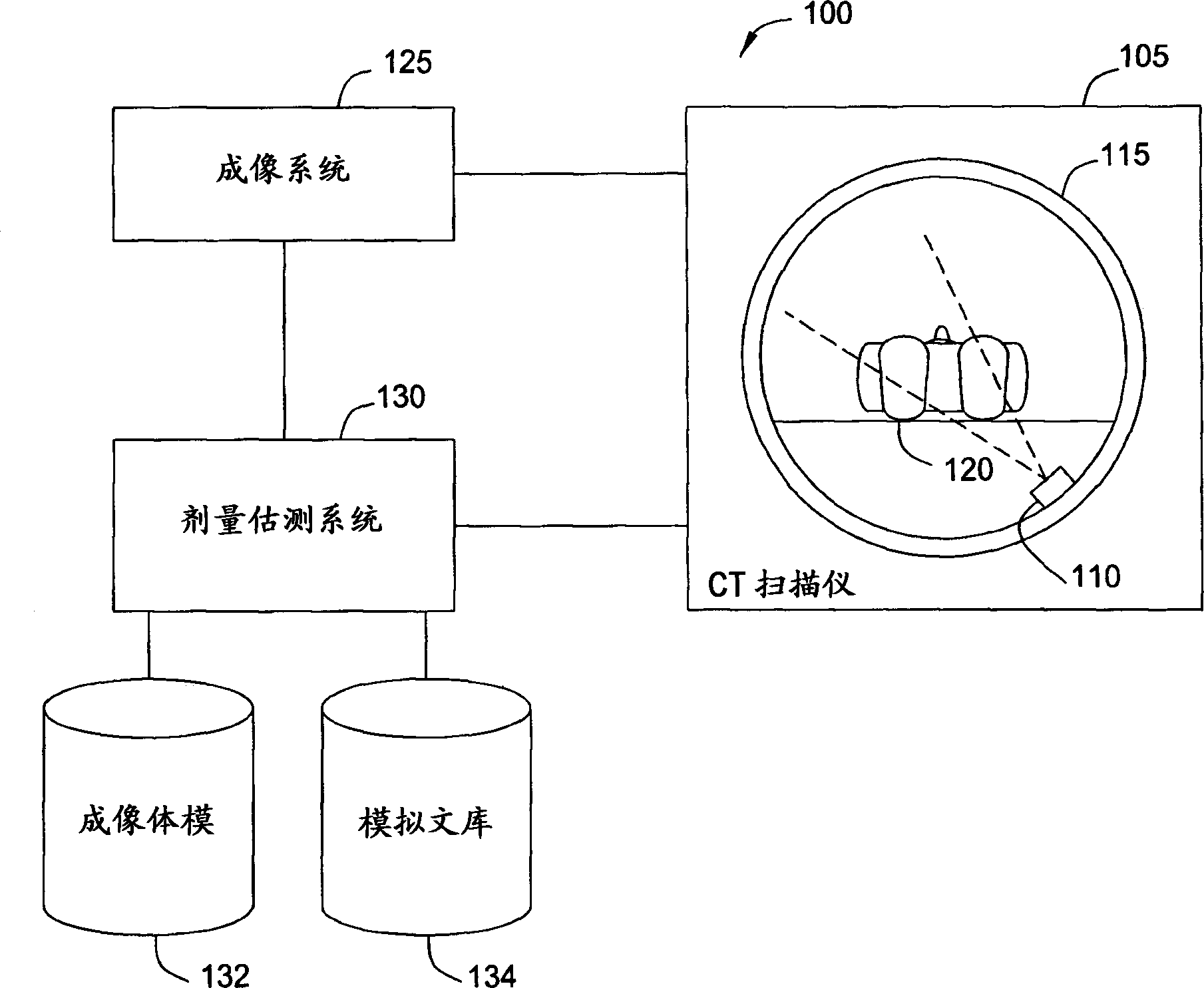
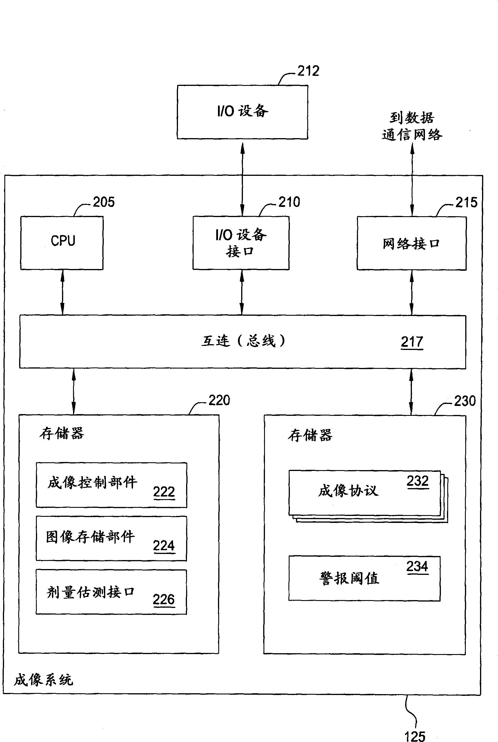
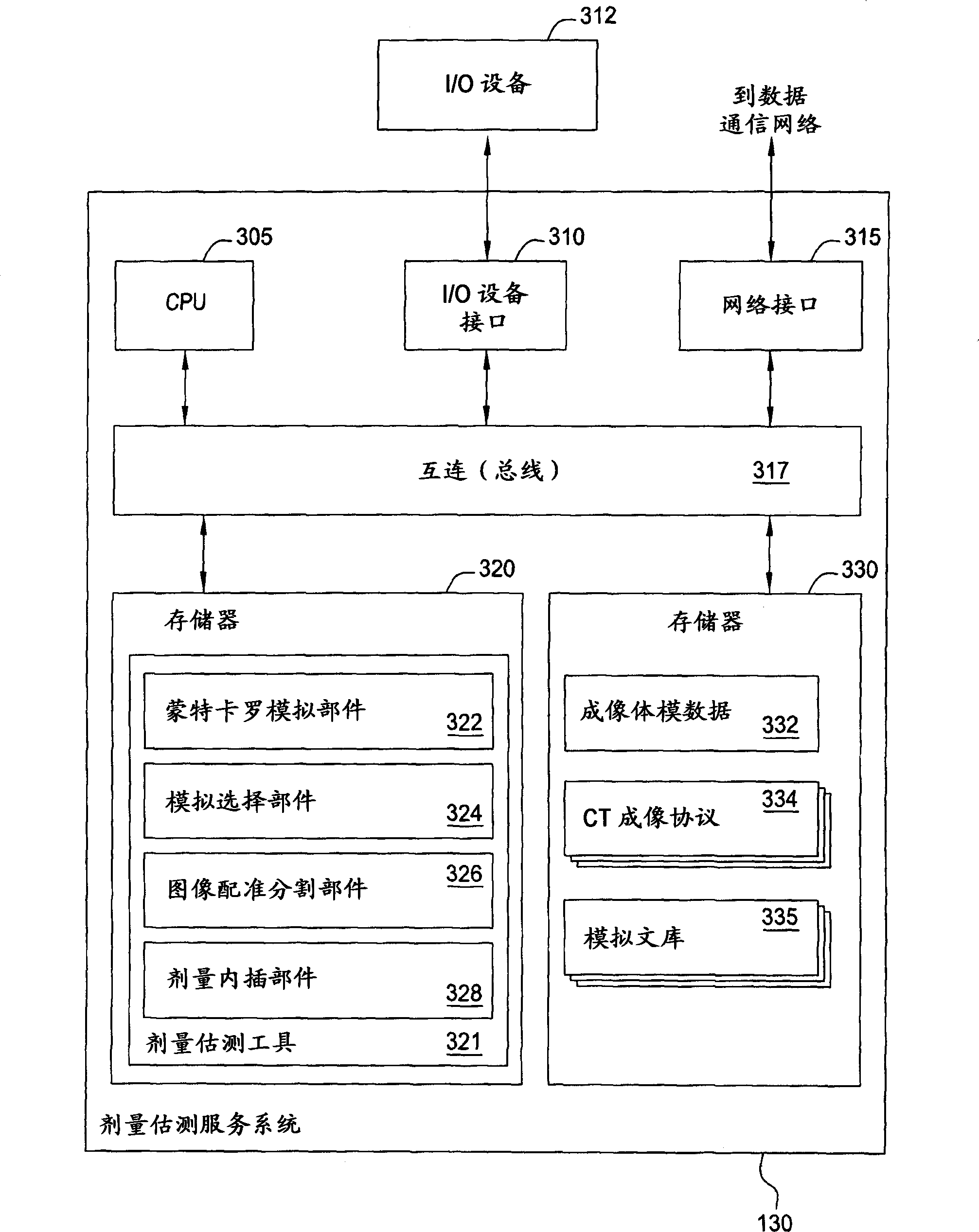
[0023] Embodiments of the invention generally relate to a scheme for estimating radiation exposure of a patient during a computerized tomography (CT) scan. More specifically, embodiments of the present invention provide efficient schemes for generating a suitable patient model for such estimation, schemes for estimating patient dose by interpolating the results of multiple simulations, and for service providers to access multiple Scheme of dose estimation services available to CT scan providers. As described in detail below, the dose management system would provide a single system for tracking radiation dose across modalities and presenting the information to practitioners in a meaningful and easily understandable form. Routine consideration of cumulative dose in custom diagnostic imaging trials can lead to a more informed decision-making process and ultimately benefit patient safety and care.

[0024] In one embodiment, a virtual imaging phantom is created to model a given p...


PUM


Abstract

Techniques are disclosed for estimating patient radiation exposure during computerized tomography (CT) scans. More specifically, embodiments of the invention provide efficient approaches for generating a suitable patient model used to make such an estimate, to approaches for estimating patient dose by interpolating the results of multiple simulations, and to approaches for a service provider to host a dose estimation service made available to multiple CT scan providers.

Description

technical field [0001] Embodiments of the invention generally relate to a scheme for estimating radiation exposure of a patient during a computerized tomography (CT) scan. Background technique [0002] As is known, CT scanning systems use ionizing radiation (X-rays) to create images of tissues, organs and other structures in the body. X-ray data from CT scans can be converted into images on a computer screen. For example, a CT scan will provide a data set used to create a three-dimensional (3D) volume corresponding to the scanned portion of the patient's body. The 3D volume is then sliced ​​to create images of body tissue at small intervals along the axis of the patient's body. Such slices may include lateral slices and transverse slices (among others), depending on the tissue or structure to be imaged. [0003] The use of CT scans and ionizing radiation for medical imaging has grown exponentially over the past decade. And modern technologies such as CT scans provide far...

Claims


Application Information

Patent Timeline
IPC IPC(8): A61B6/10A61B6/03G06T3/00G06T7/00G16H20/40G16H30/40
CPCG16H50/50A61B6/032A61B6/488A61B6/5229A61B6/542A61B6/563A61B6/583G16H20/40G16H30/40A61B6/10G06T7/0012G06T2207/10081G06V10/42G06T7/60G06T7/11G06T7/70G01T1/02G06T7/337G06T7/0014G16H30/20G06T2207/30004G06T7/00
Inventor G·库奇J·库奇
Owner BAYER HEALTHCARE LLC
Why Eureka
Try Eureka