Method for monitoring the X-ray dosage administered to a patient by a radiation source when using an X-ray device, and X-ray device

a radiation source and patient technology, applied in the direction of instruments, radiation beam directing means, diagnostic recording/measuring, etc., can solve the problem of often insufficient information, and achieve the effect of better instruments for assessing the exposure of the patien

Active Publication Date: 2013-12-17
SIEMENS HEALTHCARE GMBH
View PDF18 Cites 10 Cited by
  • Summary
  • Abstract
  • Description
  • Claims
  • Application Information

AI Technical Summary

Benefits of technology

[0019]Using the method according to the invention, it is also possible to provide a breakdown according to various parts of the X-ray spectrum. As mentioned in the introduction, in particular the soft part of the X-ray spectrum is absorbed on the skin surface. Provision can therefore be made for at least two location-dependent dosage values to be determined and shown for various radiation energies and / or radiation energy ranges. This allows the skin dosage to be broken down even more accurately. In particular, an average energy of the radiation occurring at the location on the surface of the patient can also be determined and displayed as a derived value in this case. In this case, it is particularly advantageous if a plurality of representations can be generated and, in particular, shown concurrently.
[0023]In a particularly effective embodiment of the present invention, provision can be made for the representation of the dosage value to be color-coded. Such a representation can also be referred to as a spectral representation. In this context, provision can be made for places of high skin dosage to be coded red, for example, and places of low skin dosage to be coded blue. In this way, it is possible to provide a representation which is particularly comprehensible and quick to grasp.
[0024]As indicated above, the method according to the invention allows the location-dependent dosage value (in particular therefore the total dosage value) and the representation to be established essentially in real time, meaning that the values and the representation are updated correspondingly whenever a new X-ray recording is effected. However, it is obviously also conceivable to determine a first estimate / determination of the exposure of the skin already in the context of the recording plan, by already determining and representing dosage values / total dosage values in advance. In a combined version, it is obviously also conceivable always to already take into consideration the next image that is to be recorded. Any chosen configuration is possible here, in order to ensure that the user is offered the best possible information that is needed.
[0025]In addition to the method, the present invention also relates to an X-ray device comprising in particular a radiation source which is arranged on a C-arm, a radiation detector, a patient support plate, a display device and a control device which is designed for performing the method according to the invention. Using such an X-ray device, it is therefore possible to determine and display location-dependent dosage values or values derived therefrom on the surface of a patient, either in real time and / or calculated in advance, in order thus to give the person conducting an examination a better instrument for assessing the exposure of the patient. Consequently, the user can perform a better evaluation and possibly optimize their mode of working with reference to the previously applied skin dosage, e.g. change the projection direction, invert the projection—transpose the positions of radiation source and radiation detector—or utilize low-dosage diagnostic programs where this is sufficient for the recording to be carried out.

Problems solved by technology

Disadvantageously, however, this information is often insufficient because it reveals very little about the presence of especially exposed regions on the skin of a patient, in particular.

Method used


Image

  • Method for monitoring the X-ray dosage administered to a patient by a radiation source when using an X-ray device, and X-ray device
  • Method for monitoring the X-ray dosage administered to a patient by a radiation source when using an X-ray device, and X-ray device
  • Method for monitoring the X-ray dosage administered to a patient by a radiation source when using an X-ray device, and X-ray device

Examples


Embodiment Construction

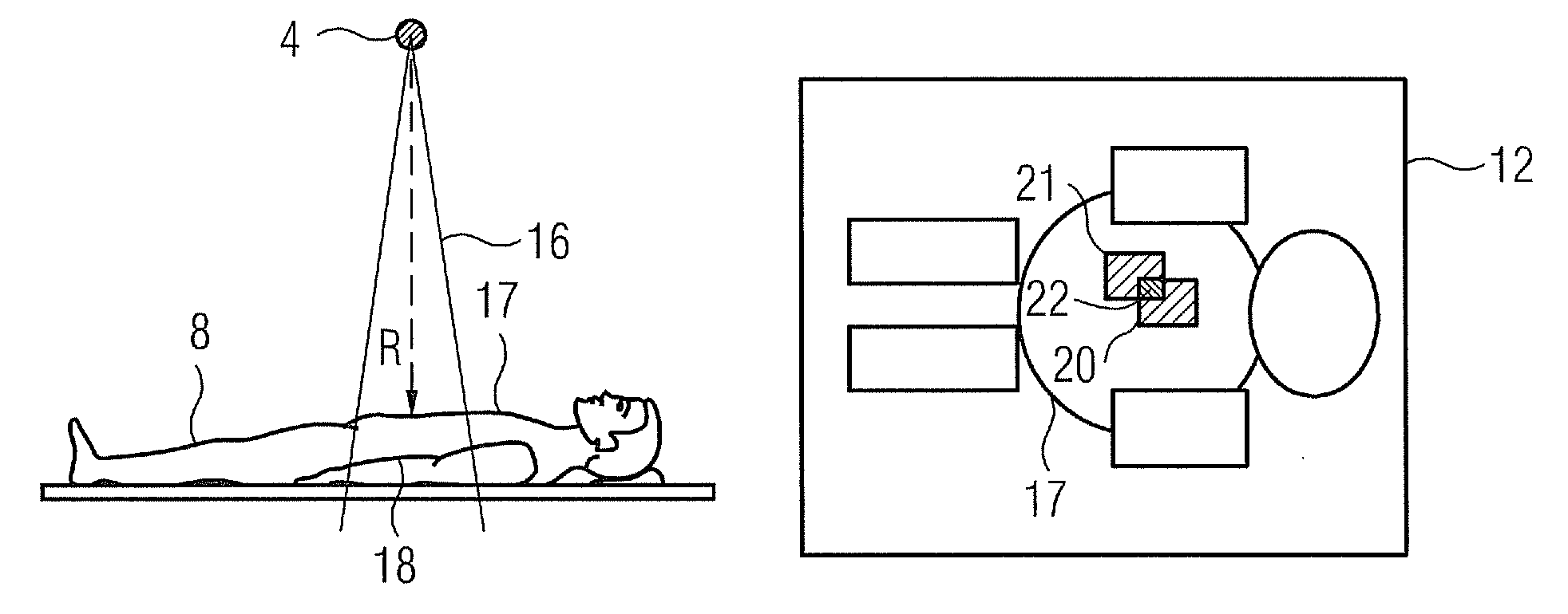
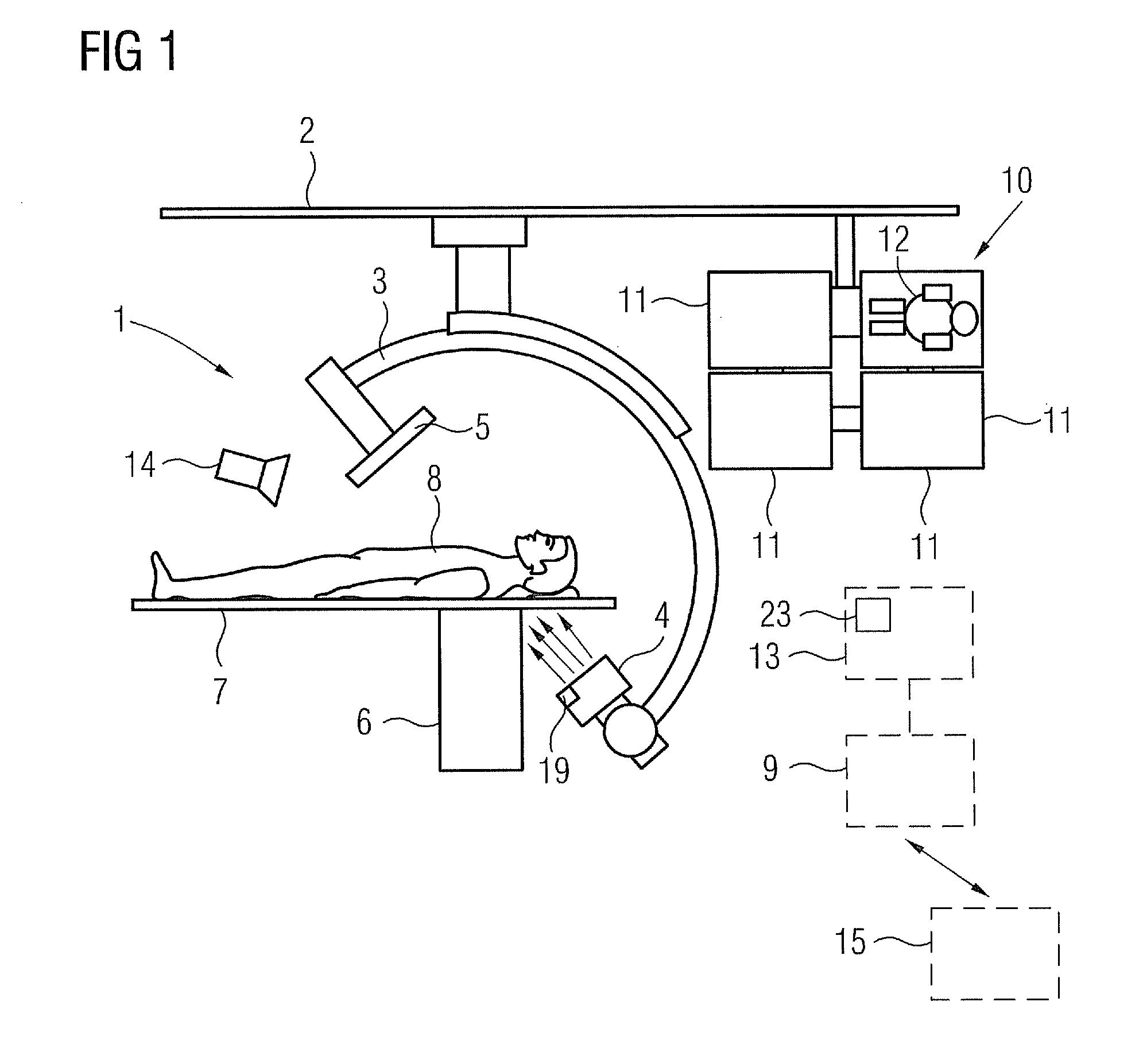
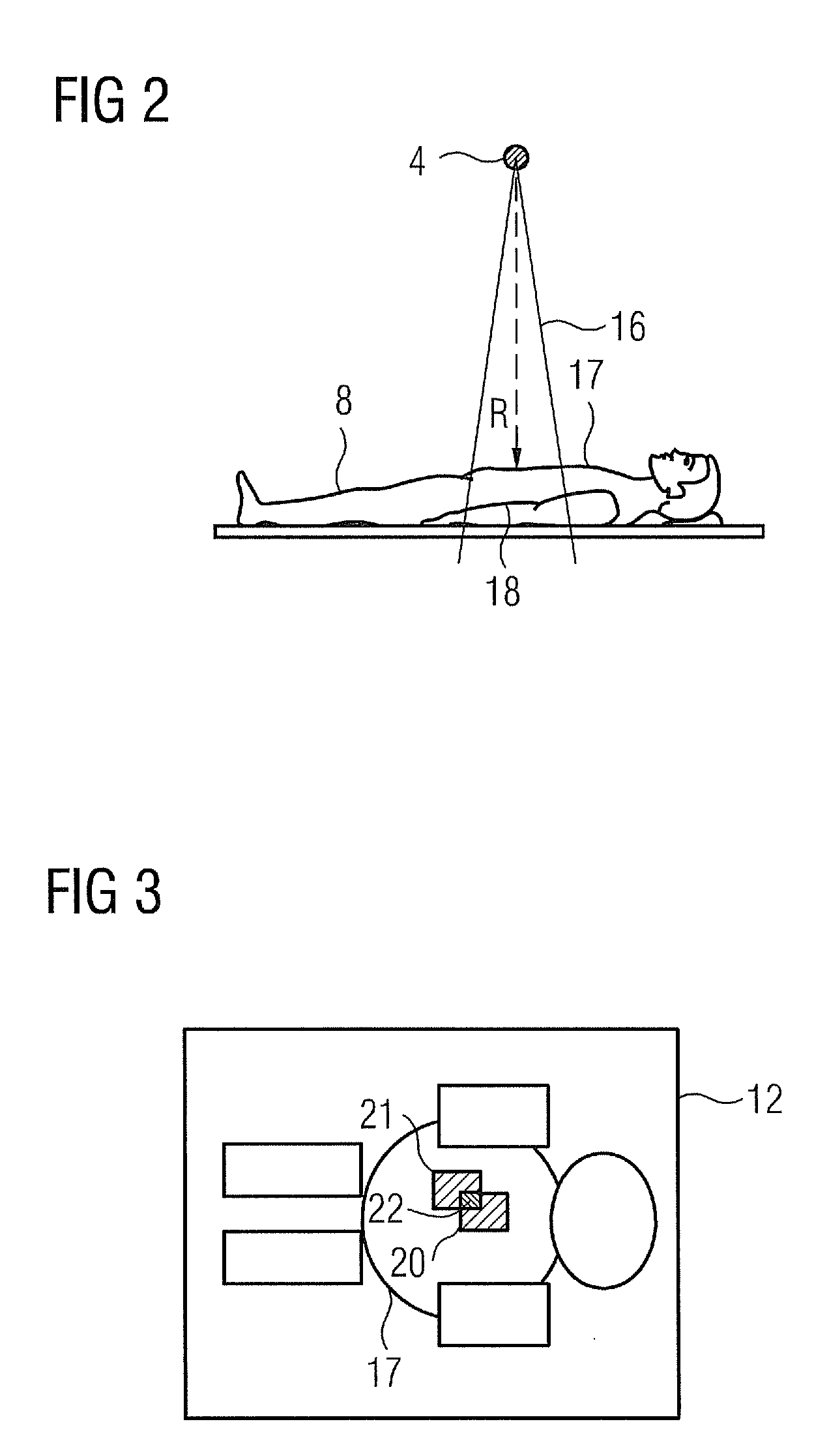
[0031]FIG. 1 shows an X-ray device 1 according to the invention, specifically a C-arm X-ray device in this case. It comprises a C-arm 3, this being attached to a ceiling 2 in the present example, on which a radiation source 4 (X-ray tube) and a radiation detector 5 (e.g. a semiconductor radiation detector) are arranged facing each other. The C-arm 3 is movable, in particular rotatable, relative to a patient couch 6 comprising a patient support plate 7 on which a patient 8 can be positioned for examination. The X-ray device 1 further comprises a control device 9 which is only shown schematically here and is designed to carry out the method according to the invention. Provision is further made for a display device 10 comprising a plurality of monitors 11, on which a representation 12 of a location-dependent dosage value on the surface of the patient 8 can be shown in addition to recorded images and / or recording parameters. Recording parameters can be entered and the components of the ...


PUM

PropertyMeasurementUnit
total currentaaaaaaaaaa
areaaaaaaaaaaa
ultrasoundaaaaaaaaaa

Abstract

A method for monitoring the X-ray dosage administered to a patient by a radiation source when using an X-ray device is proposed. The X-ray device is in particular a C-arm X-ray device. A location-dependent dosage value on the surface of the patient is determined with reference to parameters which describe the recording geometry and the radiation that is administered. The surface is described by a patient model in particular. A representation of the dosage value and / or of a value derived therefrom is displayed.

Description

CROSS REFERENCE TO RELATED APPLICATIONS[0001]This application claims priority of German application No. 10 2009 021 239.6 filed May 14, 2009, which is incorporated by reference herein in its entirety.FIELD OF THE INVENTION[0002]The invention relates to a method for monitoring the X-ray dosage administered to a patient by a radiation source when using an X-ray device, in particular a C-arm X-ray device, and an associated X-ray device.BACKGROUND OF THE INVENTION[0003]X-ray devices of various types are well known and are used for the purpose of diagnostic examination, but are also used for monitoring interventions in the context of e.g. cardiology, radiology and neurosurgery. Use is often made of C-arm X-ray devices comprising at least one C-arm, on which a radiation source and a detector are arranged facing each other, a high-voltage generator for generating the X-ray voltage of the radiation source, an imaging system including a display device, a control device, and a patient couch w...

Claims


Application Information

Patent Timeline
Patent Type & Authority Patents(United States)
IPC IPC(8): H05G1/26H05G1/64G01N23/083G01N23/04
CPCA61B6/08A61B6/4441A61B6/4464A61B6/464A61B6/542A61B6/545A61B6/589A61B5/1114
Inventor SPAHN, MARTIN
Owner SIEMENS HEALTHCARE GMBH
Why Eureka
Try Eureka