
Method for segmenting uterine fibroid ultrasound image in HIFU treatment

A technology for ultrasound images and mid-uterus, applied in the field of image processing, which can solve the problems of image noise sensitivity, excessive contraction, weak borders, etc.

Active Publication Date: 2014-05-14
WUHAN UNIV
View PDF3 Cites 7 Cited by
  • Summary
  • Abstract
  • Description
  • Claims
  • Application Information

AI Technical Summary

Problems solved by technology

However, since the gradient information is a highly localized image information, the model has two main disadvantages: sensitivity to image noise and sensitivity to initialization contours
These models have introduced local information and are more suitable for the segmentation problem of ultrasound images. They have good segmentation ability for images with non-uniform gray scale, but for those with serious low signal-to-noise ratio, low contrast, weak boundaries, etc. Ultrasound images of problematic fibroids are still prone to border leaks or missegmentation of hyperconstriction

Method used


Image

  • Method for segmenting uterine fibroid ultrasound image in HIFU treatment
  • Method for segmenting uterine fibroid ultrasound image in HIFU treatment
  • Method for segmenting uterine fibroid ultrasound image in HIFU treatment

Examples


Embodiment Construction

[0060] The present invention will be further elaborated below in conjunction with the accompanying drawings and specific embodiments.

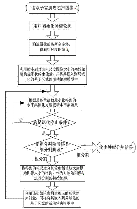
[0061] please see figure 1 , the technical solution adopted in the present invention is: a method for segmenting ultrasonic images of hysteromyoma in HIFU treatment, comprising the following steps:

[0062] Step 1: Reading Raw Image I 0 , for the original image I 0 Carry out coarse-scale segmentation, and its specific implementation includes the following sub-steps:

[0063] Step 1.1: Initialize the target contour, in the original image I 0 Draw an ellipse as the original image I 0 The initial profile C 0 , so that it can cover the original image I 0 The edge contour of the tumor in the center; the ellipse initialization is chosen because the shape of uterine fibroids is mostly similar to the ellipse shape, and it is easier to obtain a better initialization effect;

[0064] Step 1.2: Set the original image I 0 The size is M×N, accordin...


PUM


Abstract

The invention discloses a method for segmenting a uterine fibroid ultrasound image in HIFU treatment. The method comprises the implementation procedures that a tumor contour is manually initialized by a user; an image Gaussian pyramid is established to obtain a coarse scale image; shape constraint energy is established, and a local area active contour model after shape constraint is used for conducting coarse segmentation on the coarse scale image; the obtained coarse scale segmentation result is used as an initial contour for segmenting an original image, and the local area active contour model after shape constraint is used again for conducting fine segmentation on the original image; the fine segmentation result is the final tumor segmentation contour. According to the method, one new shape constraint is introduced, so that the precision for segmenting the uterine fibroid ultrasound image is improved, the problem of boundary leakage or excessive contraction caused by ultrasound image quality is solved, and the segmenting efficiency is greatly improved through implementation of a multiscale algorithm.

Description

technical field [0001] The invention relates to the technical field of image processing, and relates to a method for segmenting ultrasonic images of uterine fibroids in HIFU treatment, and a method for segmenting ultrasonic images of uterine fibroids in HIFU treatment based on a shape-constrained multi-scale local area active contour model. Background technique [0002] Uterine fibroids are a common benign tumor that plagues women. At present, high-intensity focused ultrasound (HIFU) therapy, as a new type of non-invasive surgery, has been gradually applied to the treatment of uterine fibroids due to its safety and effectiveness, reducing the pain of patients in traditional surgery. As we all know, due to its low signal-to-noise ratio and weak boundary, the accurate segmentation of ultrasound images has always been a difficult problem in the field of segmentation, and there is still no perfect solution. However, the ultrasonic images used in HIFU treatment have more serious...

Claims


Application Information

Patent Timeline
IPC IPC(8): G06T7/00
Inventor 袁志勇郑奇廖祥云
Owner WUHAN UNIV
Learn More