A method and device for segmenting images based on radionuclide imaging

A radionuclide and imaging technology, applied in the field of image processing, can solve the problem of uncertain segmentation results, and achieve the effect of reducing manual intervention, improving certainty, and avoiding subjectivity.

Active Publication Date: 2016-12-21
福建睿柠信息技术有限公司
View PDF5 Cites 0 Cited by
  • Summary
  • Abstract
  • Description
  • Claims
  • Application Information

AI Technical Summary

Problems solved by technology

[0004] In view of this, the present invention provides a segmentation method based on radionuclide imaging images, the purpose is to solve the problem of uncertain segmentation results caused by existing PET and SPECT image segmentation methods

Method used


Image

  • A method and device for segmenting images based on radionuclide imaging
  • A method and device for segmenting images based on radionuclide imaging
  • A method and device for segmenting images based on radionuclide imaging

Examples


Embodiment Construction

[0055] The following will clearly and completely describe the technical solutions in the embodiments of the present invention with reference to the accompanying drawings in the embodiments of the present invention. Obviously, the described embodiments are only some, not all, embodiments of the present invention. Based on the embodiments of the present invention, all other embodiments obtained by persons of ordinary skill in the art without making creative efforts belong to the protection scope of the present invention.

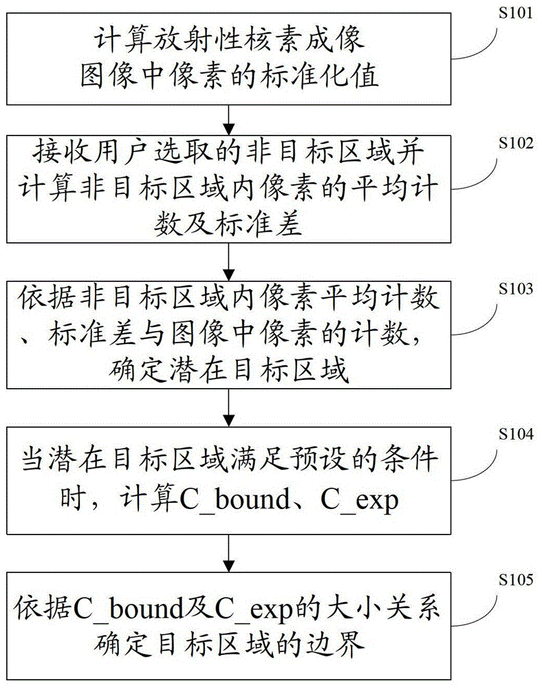
[0056] A segmentation method based on radionuclide imaging images disclosed in the embodiments of the present invention is applied to radionuclide imaging, including PET and SPECT images. The embodiments of the present invention are all described using PET images as examples, and the methods are applicable to SPECT images, the processing process is similar and will not be repeated here. Such as figure 1 As shown, the method includes:

[0057] S101: Calculate...


PUM


Abstract

Provided are a segmentation method and device based on an image of radionuclide imaging, including: determining a potential target region from an image of radionuclide imaging using a standardized value of pixels in the image of radionuclide imaging and an average standardized value and a standard deviation of pixels in a non-target region, and according to the boundary pixels and pixels within a boundary of the potential target region, and the average standardized value of the pixels of the non-target region, finally determining the boundary of a target of interest. The method or device needs to receive a non-target region selected by a user only when calculating an average standardized value and a standard deviation of pixels in the non-target region, thus significantly reducing manual intervention of a link, so as to avoid the subjectivity of segmentation results and improve the certainty of the segmentation results.

Description

technical field [0001] The invention relates to the field of image processing, in particular to a segmentation method and device based on radionuclide imaging images. Background technique [0002] Radionuclide imaging images include Positron Emission Tomography (PET) images, referred to as PET images, and Single Photon Emission Computed Tomography (SPECT) images, referred to as SPECT images, radionuclide Imaging images have a wide range of applications in the medical field. When using PET or SPECT images for medical quantitative or semi-quantitative diagnosis, it is usually necessary to segment them, that is, to separate the region of interest in the image from the background. [0003] PET or SPECT images have the characteristics of low contrast and low spatial resolution. Therefore, the existing automatic image segmentation methods cannot accurately segment the region of interest. The existing segmentation of PET or SPECT images generally consists of Experienced doctors u...

Claims


Application Information

Patent Timeline
Patent Type & Authority Patents(China)
IPC IPC(8): G06T7/00
CPCG06T7/143G06T7/11G06T2207/10104G06T2207/10108G06T2207/20036G06T2207/20104
Inventor 陈仰纯
Owner 福建睿柠信息技术有限公司
Why Eureka
Try Eureka