Method for automatically positioning standard tangent plane from ultrasonic image

An ultrasound image and automatic positioning technology, which is applied in the field of medical image processing, can solve problems such as interference, difficulty in locating standard slices, and failure to obtain results, achieving high accuracy, eliminating interference from anatomical structure detection, and overcoming the problem of position changes Effect

Active Publication Date: 2014-07-30
EDAN INSTR
View PDF2 Cites 14 Cited by
  • Summary
  • Abstract
  • Description
  • Claims
  • Application Information

AI Technical Summary

Problems solved by technology

[0004] Although existing studies have proved the feasibility of automatically positioning standard slices from two-dimensional ultrasound images, this work is still extremely challenging. For example, the automatic positioning of standard slices in the fetal abdomen is mainly due to the following reasons: First, if figure 1 As shown, only when the three key anatomical structures of gastric bubble (stomach bubble, SB), umbilical vein (umbilical vein, UV), and spine (spine, SP) exist simultaneously in an ultrasound view, this view can be defined as Fetal Abdominal Standard Plane (FASP)
However, due to the existence of artifacts, deformation of key anatomical structures, changes in image brightness, changes in fetal position, and different scanning directions, it is difficult to locate the standard slice as a whole.
Secondly, the shape of many areas in the image is very similar to the key anatomical structures, which will cause greater interference, so the detection of each anatomical structure cannot get good results

Method used


Image

  • Method for automatically positioning standard tangent plane from ultrasonic image
  • Method for automatically positioning standard tangent plane from ultrasonic image
  • Method for automatically positioning standard tangent plane from ultrasonic image

Examples


Embodiment 1

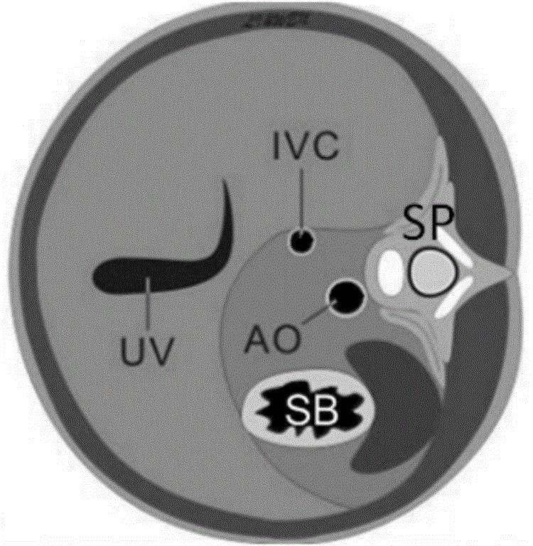
[0041] The present invention is described by taking the standard section of the fetal abdomen as an example. It can be understood that, at this time, the fetal abdominal area is the target area. Meanwhile, in this embodiment, the machine learning method adopts the random forest algorithm, of course, it is not limited to this, and other machine learning algorithms may also be used, such as: Adaboost, SVM and so on.

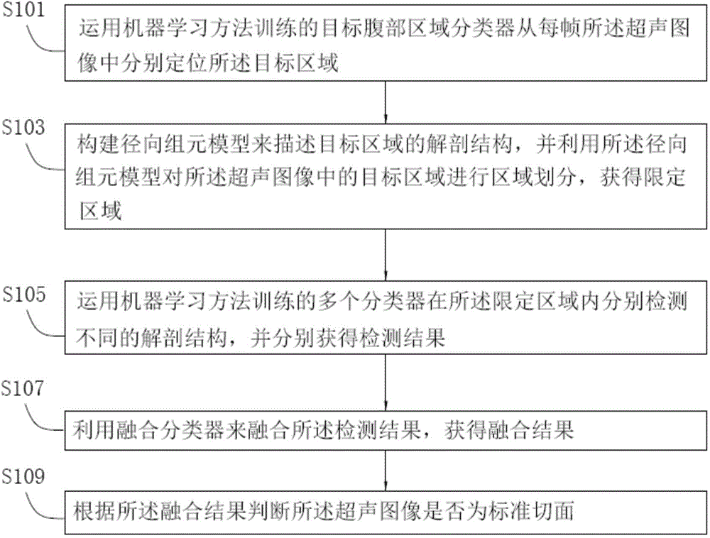
[0042] When applying the method for automatically positioning the standard section from the ultrasound image to automatically locate the standard section of the fetal abdomen, it specifically includes the following steps:

[0043] S101. Locate the abdominal region of the fetus from each frame of the ultrasound image using the abdominal region classifier trained by the random forest algorithm.

[0044] As a new type of classifier, random forests (RF) classifiers have been widely used in target detection. Compared with traditional classifiers, random forest classifie...


PUM


Abstract

The invention brings forward a method for automatically positioning a standard tangent plane from an ultrasonic image. The method comprises the following steps: S103, constructing a radial component module to describe the anatomical structure of a target area, performing area dividing on the target area in the ultrasonic image by use of the radial component module, and obtaining a limitation area; S105, respectively detecting different anatomical structures in the limitation area by use of multiple classifiers trained by a machine learning method, and respectively obtaining detection results; S107, fusing the detection results by use of a fusion classier, and obtaining a fusion result; and S109, according to the fusion result, determining whether the ultrasonic image is the standard tangent plane. The method provided by the invention can effectively improve the detection accuracy.

Description

technical field [0001] The invention relates to medical image processing technology, in particular to a method for automatically locating a standard slice from an ultrasonic image based on radial components, which can effectively improve detection accuracy. Background technique [0002] Ultrasound (Ultrasound, US) is widely used in routine clinical examination and prenatal diagnosis. Routine ultrasonography is divided into three steps: probe scanning, selection of standard section and diagnosis. Among them, the selection of the standard section is very important for the accurate measurement and diagnosis of the physiological parameters of the examination object, such as the fetus. However, this work requires extensive clinical experience and an in-depth understanding of human anatomy, so it is extremely challenging for inexperienced clinicians, and it is time-consuming and complicated even for experienced doctors. This is also often considered an inherent disadvantage of u...

Claims


Application Information

Patent Timeline
IPC IPC(8): G06K9/62
Inventor 倪东杨鑫李胜利汪天富陈思平
Owner EDAN INSTR
Why Eureka
Try Eureka