Preparation method of three-dimensional contraction model for constructing artificial blood vessel model

An artificial blood vessel and model technology, applied in the field of blood vessel models, can solve the problems of slow cell proliferation and low mechanical strength, and achieve the effects of fast growth, short model making cycle and strong mechanical strength

Active Publication Date: 2014-10-15
WENZHOU MEDICAL UNIV
View PDF5 Cites 2 Cited by
  • Summary
  • Abstract
  • Description
  • Claims
  • Application Information

AI Technical Summary

Problems solved by technology

[0004] Aiming at the shortcomings of slow cell proliferation and low mechanical strength in the current three-dimensional contraction model production process, the present invention provides a method for preparing a three-dimensional contraction model for constructing an artificial blood vessel model

Method used


Image

  • Preparation method of three-dimensional contraction model for constructing artificial blood vessel model
  • Preparation method of three-dimensional contraction model for constructing artificial blood vessel model
  • Preparation method of three-dimensional contraction model for constructing artificial blood vessel model

Examples


Embodiment 1

[0035] A method for preparing a three-dimensional contraction model for constructing an artificial blood vessel model, comprising the steps of:

[0036]Step (1), the preparation of collagen cell mixture: the preparation method of 2ml collagen cell mixture is that 467 μl concentration is the type I collagen of 8.56mg / ml, 200 μl 10×phosphate buffer saline, 10.7 μl concentration is the sodium hydroxide of 1M After mixing evenly, add fetal bovine serum, DMEM medium and 20*10 6 The total volume of the mixed solution of each myofibroblast is 2ml, and the volume ratio of the fetal bovine serum and DMEM medium is 1: 9, blow gently with a pipette until the mixture is even, to obtain final product;

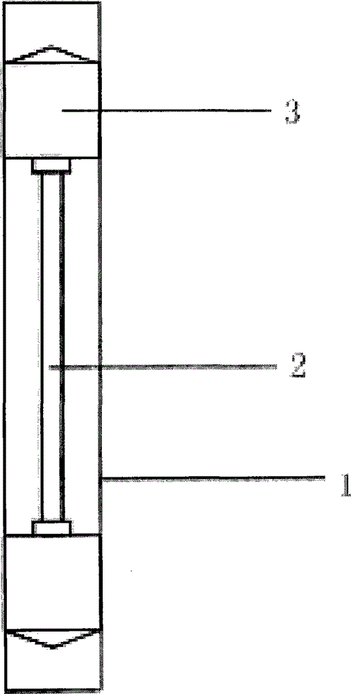
[0037] Step (2), the cross-linking solidification of collagen cell mixture: as figure 1 As shown, the Teflon cylindrical tube has a built-in glass tube inner core, and the upper and lower ends are closed with rubber stoppers to make a cylindrical model device; inject 2 ml of the collagen c...

Embodiment 2

[0040] A method for preparing a three-dimensional contraction model for constructing an artificial blood vessel model, comprising the steps of:

[0041] Step (1), preparation of collagen cell mixture: the preparation method of 2ml collagen cell mixture is to mix 3.9 μl of type I collagen, 200 μl of 10× phosphate buffer saline, and 10.5 μl of sodium hydroxide with a concentration of 1M, and then add fetal Bovine serum, DMEM medium and 19*10 6 The total volume of the mixed solution of each myofibroblast is 2ml, and the volume ratio of the fetal bovine serum and DMEM medium is 1: 9, blow gently with a pipette until the mixture is even, to obtain final product;

[0042] Step (2), the cross-linking solidification of collagen cell mixture: as figure 1 As shown, the Teflon cylindrical tube has a built-in glass tube inner core, and the upper and lower ends are closed with rubber stoppers to make a cylindrical model device; inject 2 ml of the collagen cell mixture obtained in step (1)...

Embodiment 3

[0045] A method for preparing a three-dimensional contraction model for constructing an artificial blood vessel model, comprising the steps of:

[0046] Step (1), preparation of collagen cell mixture: the preparation method of 2ml collagen cell mixture is to mix 4.1g type I collagen, 200 μl 10× phosphate buffer saline, 10.9 μl concentration of 1M sodium hydroxide, and add Fetal bovine serum, DMEM medium and 21*10 6 The total volume of the mixed solution of each myofibroblast is 2ml, and the volume ratio of the fetal bovine serum and DMEM medium is 1: 9, blow gently with a pipette until the mixture is even, to obtain final product;

[0047] Step (2), the cross-linking solidification of collagen cell mixture: as figure 1 As shown, the Teflon cylindrical tube has a built-in glass tube inner core, and the upper and lower ends are closed with rubber stoppers to make a cylindrical model device; inject 2 ml of the collagen cell mixture obtained in step (1) into the cylindrical model...


PUM

PropertyMeasurementUnit
diameteraaaaaaaaaa
diameteraaaaaaaaaa

Abstract

The invention relates to a preparation method of a three-dimensional contraction model for constructing an artificial blood vessel model, and belongs to the technical field of blood vessel models. The method comprises the steps of preparing a collencyte mixed solution, crosslinking and solidifying the collencyte mixed solution, and preparing the three-dimensional contraction model. The prepared three-dimensional contraction model serves as an improved contraction model, adopts myofibroblast IMR-90, grows faster, and is shorter in preparation cycle; auxiliary contraction factors U0126 and TGF-beta are added to stimulate the myofibroblast, so that the three-dimensional contraction model expresses contractive protein, has better contractility, and further has higher mechanical strength; and the prepared three-dimensional contraction model can provide a better and faster contraction model for scientific research, and can provide reference for later clinical application.

Description

technical field [0001] The invention belongs to the technical field of blood vessel models, and in particular relates to a method for preparing a three-dimensional contraction model for constructing an artificial blood vessel model. Background technique [0002] At present, the three-dimensional shrinkage model is mostly used in scientific research or bioengineering to construct artificial blood vessel models, and even used in clinic as a substitute for blood vessels. Clinically, although relatively mature biomaterials have been used to replace larger blood vessels, there is still no ideal substitute for small arteries such as the coronary arteries of the heart. Although native blood vessels can be an option, many patients have vascular diseases such as atherosclerosis, which makes it possible to find suitable biological replacement products as a treatment method. Currently available methods for making three-dimensional shrinkage models for scientific research have shortcom...

Claims


Application Information

Patent Timeline
Patent Type & Authority Applications(China)
IPC IPC(8): A61L27/38
Inventor 祝建洪赖俊媚张雄
Owner WENZHOU MEDICAL UNIV
Why Eureka
Try Eureka