X-ray tomography method and X-ray tomography system

A tomography and X-ray technology, applied in the field of X-ray tomography methods and systems, can solve the problems of reduced spatial resolution of the scanning system, large focal spot blur area, patient movement, etc., to save scanning time and improve spatial resolution , Improve the effect of scanning speed

Active Publication Date: 2015-02-04
SHENZHEN INST OF ADVANCED TECH CHINESE ACAD OF SCI
View PDF6 Cites 9 Cited by
  • Summary
  • Abstract
  • Description
  • Claims
  • Application Information

AI Technical Summary

Problems solved by technology

However, the higher the scanning speed, the greater the distance the X-ray tube moves within the exposure time window, and the resulting blurred area of ​​the focal spot is also larger, thus reducing the spatial resol

Method used


Image

  • X-ray tomography method and X-ray tomography system
  • X-ray tomography method and X-ray tomography system
  • X-ray tomography method and X-ray tomography system

Examples


Embodiment Construction

[0025] In order to make the object, technical solution and advantages of the present invention clearer, the present invention will be further described in detail below in conjunction with the accompanying drawings and embodiments. It should be understood that the specific embodiments described here are only used to explain the present invention, not to limit the present invention.

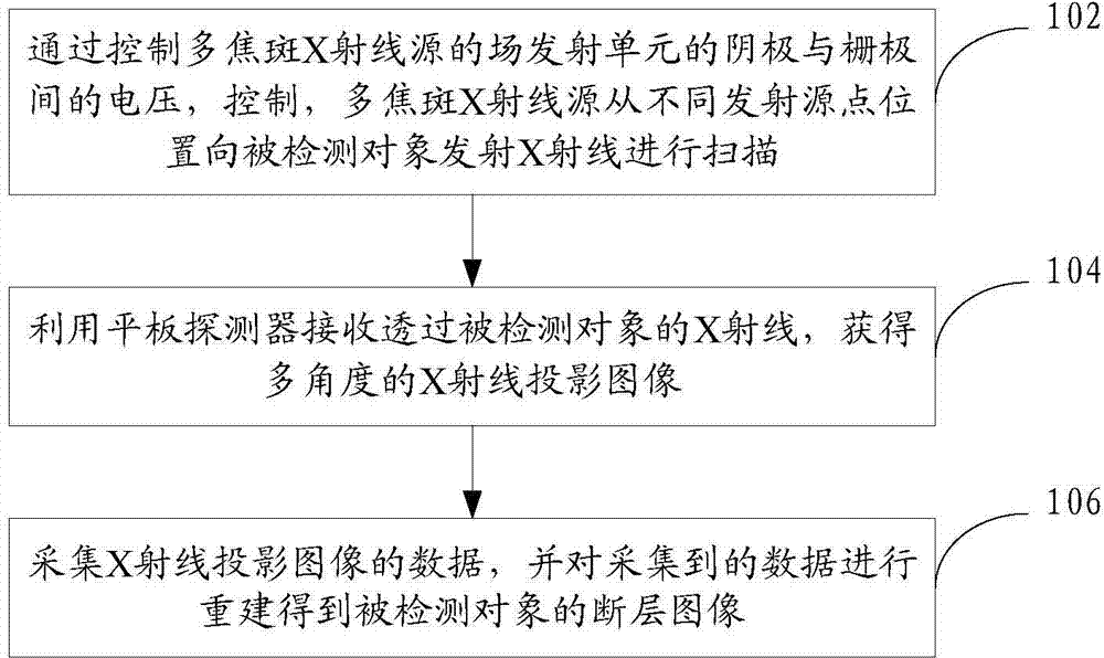
[0026] In one embodiment, such as figure 1 As shown, a method for X-ray tomography is provided, the method comprising:

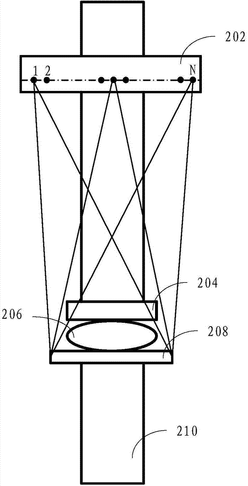
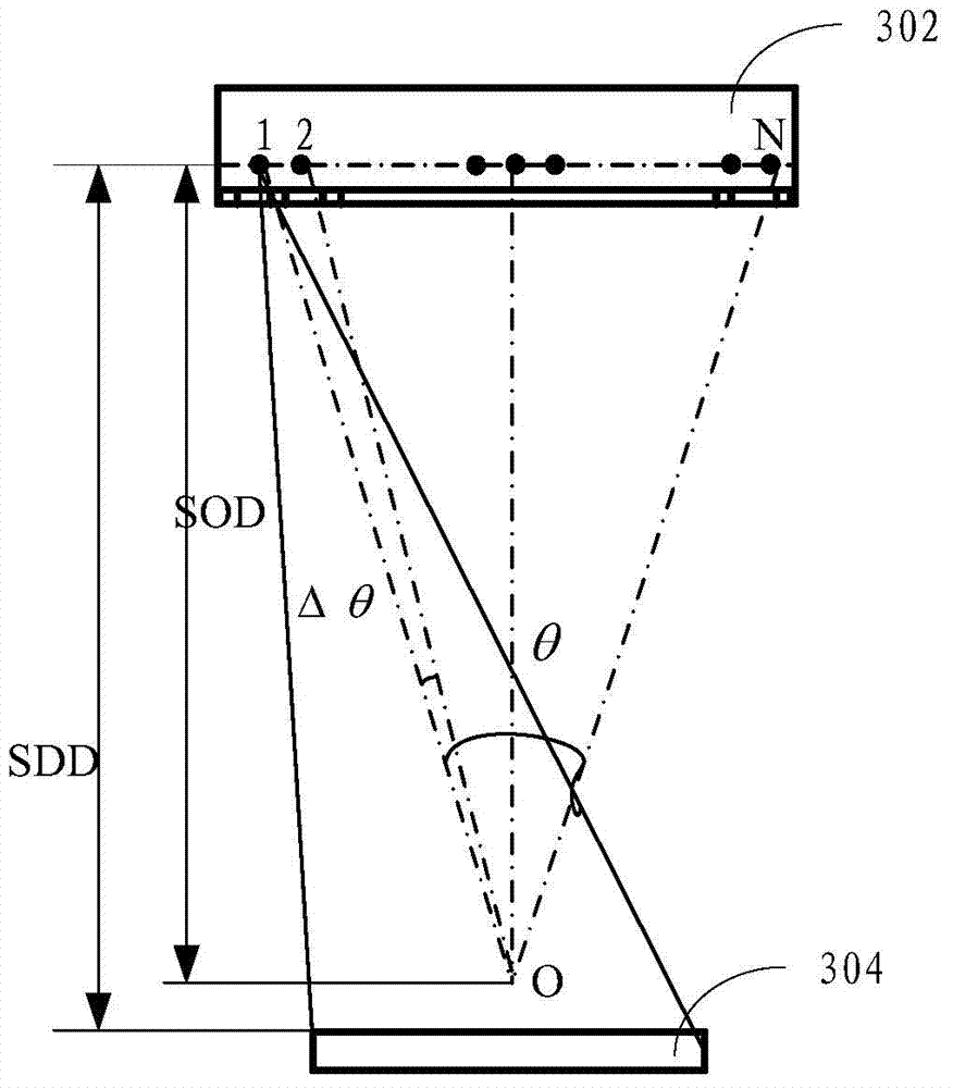
[0027] Step 102, by controlling the voltage between the cathode and grid of the field emission unit of the multi-focal X-ray source, the multi-focal X-ray source is controlled to emit X-rays from different emission source point positions to the detected object for scanning.

[0028] The multi-focal spot X-ray source is a plurality of field emission units arranged in a linear array, specifically, a linear array multi-focal spot carbon nanotube X-ray source can be used. The field...


PUM


Abstract

The invention provides an X-ray tomography method, which comprises the following steps: by controlling voltage between the cathode and the grid of a field emission unit of a multi-focal spot X-ray source, controlling the multi-focal spot X-ray source to emit X-rays to a detected object from different emission source positions to perform scanning; receiving the X-rays transmitting the detected object by utilizing a flat panel detector to obtain X-ray projected images at multiple angles; collecting data of the X-ray projected images, and performing reconstruction on the collected data to obtain the faulted image of the detected object. By adopting the method, not only can the spatial resolution be improved but also the scanning speed can be increased; moreover, the invention also provides an X-ray tomography system.

Description

technical field [0001] The present invention relates to the technical field of X-ray tomography, in particular to an X-ray tomography method and system. Background technique [0002] Breast cancer is one of the most common diseases that endanger women's health. Mammography X-ray examination is currently the most routine method for early screening of breast cancer. Since the X-ray inspection of mammography film obtains overlapping two-dimensional images, the information in the depth direction is lacking, resulting in more false positives or false negatives. As a new imaging technique, X-ray tomography can obtain three-dimensional image information, which can effectively avoid misdiagnosis caused by overlapping tissues. [0003] In an existing X-ray tomography system, in order to perform multi-view X-ray scanning, an X-ray tube is installed on a rotating frame, and the X-ray tube is moved along a preset arc. When the X-ray tube works in the intermittent motion mode, the X-ra...

Claims


Application Information

Patent Timeline
IPC IPC(8): A61B6/00
Inventor 桂建保陈垚郑海荣张成祥洪序达
Owner SHENZHEN INST OF ADVANCED TECH CHINESE ACAD OF SCI
Why Eureka
Try Eureka