A device for fusion and fixation of pet, ct and mr images of experimental animals

An image fusion, experimental animal technology, applied in the direction of animal restraint equipment, applications, veterinary instruments, etc., can solve the problems of research, no fixed identification site, single function, etc., achieve easy assembly and operation, important application value, The effect of ensuring safety

Active Publication Date: 2017-02-08
THE SECOND HOSPITAL OF TIANJIN MEDICAL UNIV
View PDF6 Cites 0 Cited by
  • Summary
  • Abstract
  • Description
  • Claims
  • Application Information

AI Technical Summary

Problems solved by technology

Therefore, medical image fusion has become a hot spot in modern medical research, but the image fusion method is still immature, and it cannot be applied to human body for research. Only more data can be obtained through animal experiments to find the optimal image fusion method
[0005] However, at present, there are many kinds of devices used for immobilizing experimental animals, but their functions are single, and their materials are mostly various metal products, which are difficult to be compatible with a variety of imaging equipment. Moreover, there are no fixed identification points on experimental animals or on the immobilizing devices, which cannot realize the immobilization of experimental animals. image fusion

Method used


Image

  • A device for fusion and fixation of pet, ct and mr images of experimental animals
  • A device for fusion and fixation of pet, ct and mr images of experimental animals

Examples


Embodiment Construction

[0018] The technical solutions of the present invention will be further described in detail below in conjunction with the accompanying drawings and specific embodiments.

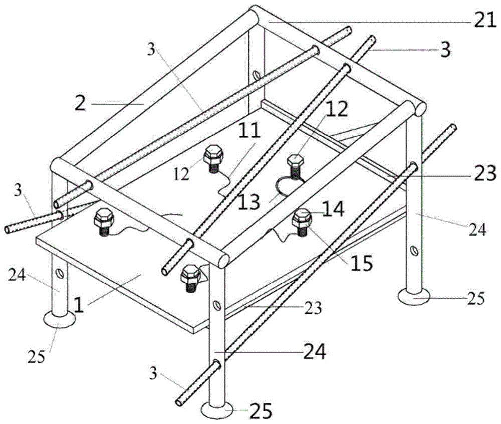
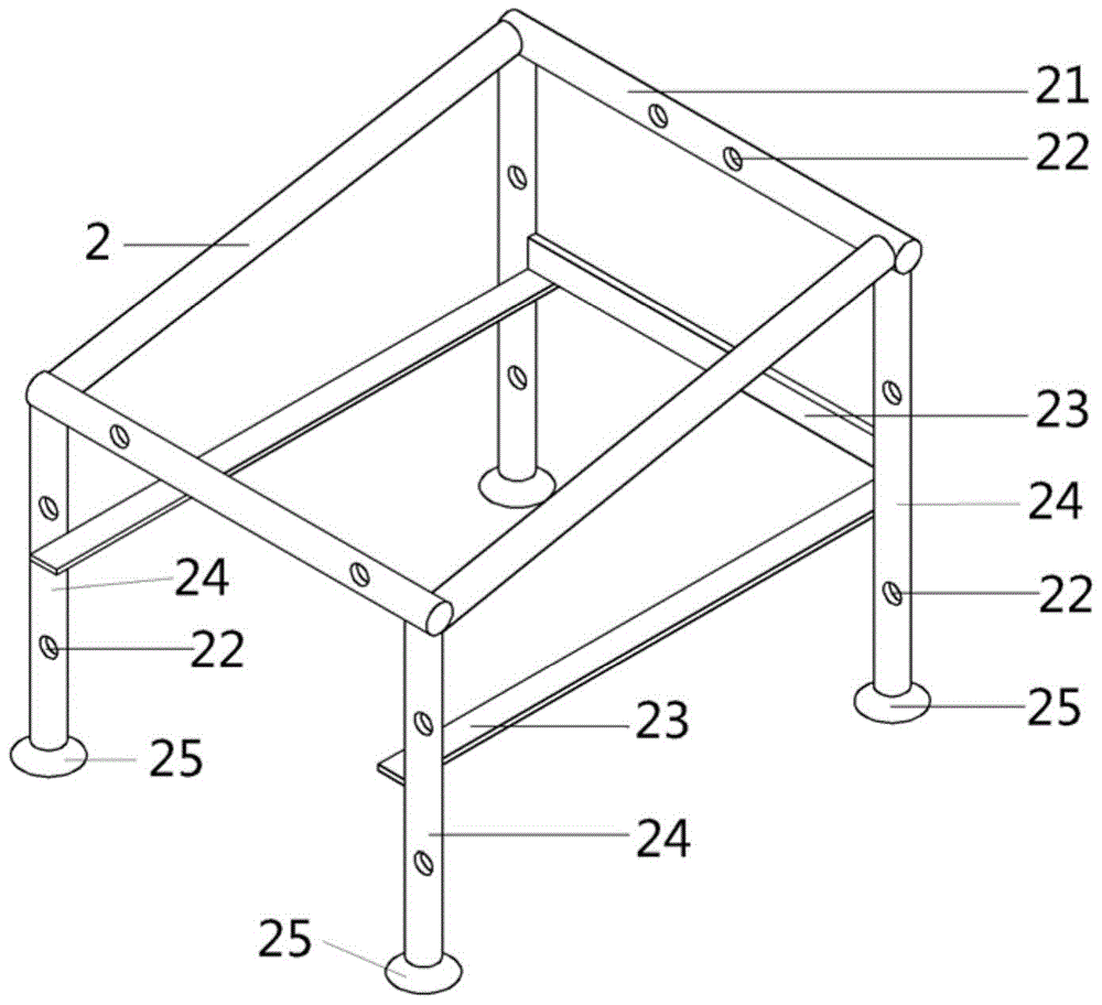
[0019] like figure 1 and figure 2 As shown, the present invention is used for experimental animal PET, CT and MR image fusion and fixation device, comprises fixed frame 2, support plate 1 and three sets of positioning rods 3, and described fixed frame 2 is made of top support frame 21 and a plurality of The support leg 24 is formed, the support leg 24 is provided with a support 23 , the support plate 1 is carried on the support 23 , and the bottom of the support leg 24 is provided with a base 25 .

[0020] The support plate 1 is provided with a plurality of positioning screws 12, one of which is fixed with an elastic rope 13, and the rest of the positioning screws 12 are respectively fixed with a fixed rope 11 and a fastening nut 15.

[0021] The positioning rod 3 is composed of a hollow tube and a plug, ...


PUM


Abstract

The invention discloses a device for PET-CT-MR image fusion and fixation of an experimental animal. The device comprises a fixed frame, a support plate and three sets of positioning rods, wherein a plurality of positioning screws are arranged on the support plate, an elastic rope is arranged on one positioning screw, and fixing ropes and clamp nuts are arranged on the other positioning screws; the three sets of positioning rods have the same number (at least three) of positioning rods; one set of positioning rods is fixed on the fixed frame simultaneously, wherein at least two positioning rods are inclined relative to each other; the positioning rods in each set are filled with PET, CT and MR photographic developers respectively, the positioning rods can be changed to add positioning information to three different image data of the experimental animal, namely PET image data, CT image data and MR image data, and accurate fusion of any more than two scan images is realized by an external feature point extraction method. The device is simple and convenient to operate, can be used repeatedly, guarantees the safety of the animal and workers, meanwhile provides a basis for deep study on image fusion methods and provides more effective, accurate and visual aid for scientific research and clinic and translation thereof.

Description

technical field [0001] The present invention relates to an auxiliary device for the integration of experimental animal fixation and experimental animal image fusion, in particular to a small animal PET, CT and MR image fusion and fixation device, which is used for experimental animal fixation and PET, CT and MR image fusion provides a basis for animal experiments and clinical transformation of image fusion technology. Background technique [0002] Experimental animals, such as rats and rabbits, are widely used as experimental objects in various medical experiments and scientific research, and are one of the more commonly used animal models in modern medical laboratories. However, rodents will resist any unconditioned stimulation, and even cause unnecessary harm to the experimenters, so it is particularly important to fix the experimental animals during the experiment. [0003] In recent years, small animal in vivo imaging has been widely used in small animal disease model r...

Claims


Application Information

Patent Timeline
Patent Type & Authority Patents(China)
IPC IPC(8): A61B6/03A61B5/055A61D3/00
Inventor 赵阳张雪宁彭景贾荣荣张骐吴梦琳郭琪李亮励贺文
Owner THE SECOND HOSPITAL OF TIANJIN MEDICAL UNIV
Why Eureka
Try Eureka