
Automatic segmentation method for retina serous pigment epithelial layer detachment

A technology for automatic segmentation of pigment epithelium, applied in medical image processing and analysis, and computer vision, can solve problems such as algorithm failure, segmentation error, and failure to provide quantitative information on lesion areas, and achieve the effect of improving accuracy

Active Publication Date: 2015-04-29
广州比格威医疗科技有限公司
View PDF2 Cites 8 Cited by
  • Summary
  • Abstract
  • Description
  • Claims
  • Application Information

AI Technical Summary

Problems solved by technology

[0003] The current automatic retinal image segmentation algorithm has the following defects: (1) Most of the algorithms are two-dimensional algorithms, that is, they are segmented independently in each slice image (x-z plane image, called transverse scanning image). Make full use of three-dimensional context information, and are more susceptible to image noise or artifacts, resulting in segmentation errors
(2) Most of the existing retinal tissue hierarchical segmentation algorithms are designed for normal retina. When the retinal tissue is deformed due to lesions, these algorithms will fail
[0004] (3) Most of the existing retinal lesion area segmentation algorithms only qualitatively analyze the lesion area, and do not provide accurate quantitative information about the lesion area, such as shape, size and location, etc.
Therefore, the accuracy of diagnosis and treatment cannot be improved

Method used


Image

  • Automatic segmentation method for retina serous pigment epithelial layer detachment
  • Automatic segmentation method for retina serous pigment epithelial layer detachment
  • Automatic segmentation method for retina serous pigment epithelial layer detachment

Examples


Embodiment Construction

[0028] The design will be further described below in conjunction with the accompanying drawings of the description.

[0029] The automatic segmentation method of retinal serous pigment epithelial layer detachment is characterized in that: comprising the following steps:

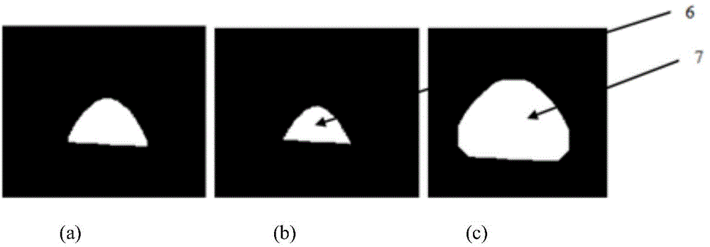
[0030] a. Preprocessing: input the three-dimensional retinal image acquired by the optical coherence tomography eye imager into the computer, and use the curve anisotropic diffusion filtering method to denoise the image of retinal serous pigment epithelial detachment; b. Automatic segmentation: first, use The image of retinal serous pigment epithelium detachment is layered by graph search algorithm (stratification refers to dividing the retinal image into different surfaces), and the initial segmentation result of the pigment epithelium detachment area is obtained; then, the foreground seed point is obtained using a mathematical morphology algorithm , background seed points; finally, use the graph cut algorit...


PUM


Abstract

The invention provides an automatic segmentation method for retina serous pigment epithelial layer detachment. The method comprises the following steps: a, pretreatment: inputting a three-dimensional retinal image obtained by an optical coherence tomography eye imager into a computer, and de-noising an image detached from a retina serous pigment epithelial layer by using a curve anisotropic diffusion filtering method; b, automatic segmentation: layering the image detached from the retina serous pigment epithelial layer by using a graph search algorithm, so as to obtain an initial segmented result; obtaining foreground and background seed points according to the initial segmented result by using a mathematical morphology algorithm; automatically segmenting a retina serous pigment epithelium detachment region by using a graph cut algorithm; c, post-treatment: optimizing the automatic segmentation result by using the mathematical morphology algorithm. According to the invention, the graph search algorithm, the graph cut algorithm and the mathematical morphology algorithm are effectively combined, so that the automatic segmentation of the retina serous pigment epithelial layer detachment region is realized.

Description

technical field [0001] The design relates to an automatic segmentation method for retinal serous pigment epithelium detachment, and belongs to the technical fields of computer vision, medical image processing and analysis. Background technique [0002] Retinal serous pigment epithelial detachment may be caused by a variety of choroidal or retinal diseases, such as age-related macular degeneration, polypoidal choroidal vasculopathy, central serous chorioretinopathy, uveitis, etc. Since retinal serous pigment epithelial detachment often results in loss of central vision, automatic segmentation of retinal serous pigment epithelial detachment has important clinical implications. [0003] The current automatic retinal image segmentation algorithm has the following defects: (1) Most of the algorithms are two-dimensional algorithms, that is, they are segmented independently in each slice image (x-z plane image, called transverse scanning image). Making full use of 3D context infor...

Claims


Application Information

Patent Timeline
IPC IPC(8): G06T7/00A61B3/12A61B3/14
CPCA61B3/1225G06T7/0012G06T7/11G06T2207/10024G06T2207/30041
Inventor 陈新建孙助力石霏
Owner 广州比格威医疗科技有限公司
Learn More