Two-dimensional and three-dimensional medical image registration method

A medical image, three-dimensional technology, applied in the field of medical imaging, can solve the problems of CT images that cannot guide treatment quickly and accurately, and difficult to register, and achieve the effect of quickly and accurately guiding treatment and registration.

Inactive Publication Date: 2015-05-20
INST OF AUTOMATION CHINESE ACAD OF SCI
View PDF4 Cites 36 Cited by
  • Summary
  • Abstract
  • Description
  • Claims
  • Application Information

AI Technical Summary

Problems solved by technology

[0002] In order to realize Image Guided Therapy (IGRT), Image Guided Radiosurgery (IGRS) and Image Guided Minimally Invasive Therapy (IGMIT), the existing surgical lesion localization process mainly includes: Three-dimensional (Three Dimension, 3D) computed tomography (CT) imaging (Computed Tomography, CT) was performed before operation; two-dimensional (Two-Dimension, 2D) X-ray images of the lesion were scanned in real time during the operation, and CT images were obtained according to The ray projection algorithm generates 2D digitally reconstructed radio-graphs (Digitally Reconstructed Radio-graphs, DRR). By measuring the similarity between the DRR and the X-ray image, the CT image and the X-ray image are registered to obtain the lesion (X-ray image) currently in the CT image. Accurate position and direction in the image, and then guide the treatment in real time. This method needs to consume a long time to generate a large number of DRRs for registration when the registration accuracy is high, so it is difficult to achieve fast and accurate registration between CT and X-ray. Accurate, so that CT images can not quickly and accurately guide the treatment

Method used


Image

  • Two-dimensional and three-dimensional medical image registration method
  • Two-dimensional and three-dimensional medical image registration method
  • Two-dimensional and three-dimensional medical image registration method

Examples


Embodiment Construction

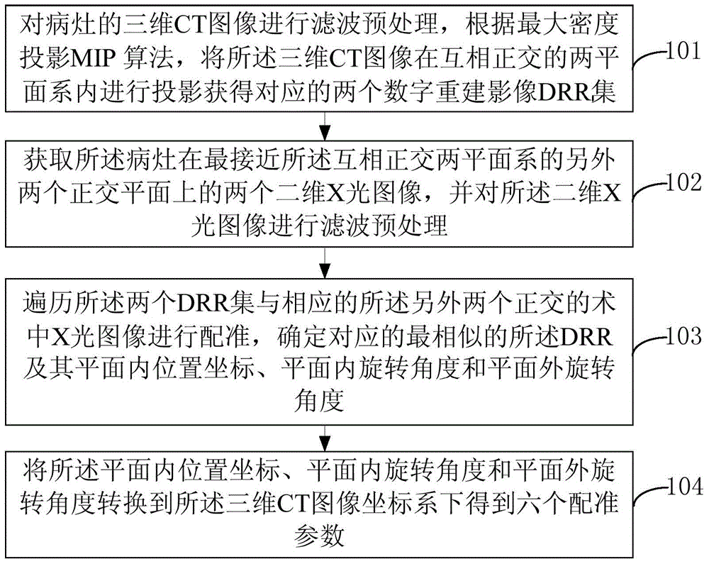
[0016] figure 1 is a flow chart of Embodiment 1 of a two-dimensional and three-dimensional medical image registration method of the present invention, such as figure 1 As shown, a two-dimensional and three-dimensional medical image registration method of the present invention includes:

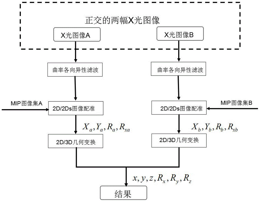
[0017] S101. Perform filtering and preprocessing on the three-dimensional CT image of the lesion, and project the three-dimensional CT image in a mutually orthogonal two-plane system according to a maximum density projection algorithm to obtain two corresponding digital reconstructed image DRR sets;

[0018] The three-dimensional CT image can also be replaced with CT, MRI, PER or 3DRA images; preferably, the x-axis is the longitudinal axis of the three-dimensional CT image of the human body, and the three-dimensional CT image is generated before surgery, that is, in the surgical planning stage. Computed tomography imaging CT images;

[0019] Preferably, the filtering preprocessing is a curva...


PUM


Abstract

The invention provides a two-dimensional and three-dimensional medical image registration method, which can quickly carry out registration on an intra-operative two-dimensional X-ray image and a preoperative three-dimensional CT (Computed Tomography) image. The two-dimensional and three-dimensional medical image registration method comprises the following steps: carrying out filtering preprocessing on the three-dimensional CT image, projecting the three-dimensional CT image in two mutually-orthogonal plane systems to obtain two corresponding digitally reconstructed radiograph (DRR) sets according to a maximum intensity projection (MIP) algorithm; obtaining two two-dimensional X-ray images on another two orthogonal planes, and carrying out the filtering preprocessing on the two-dimensional X-ray image; carrying out traversal on the two DRR sets, and carrying out registration on the two DRR sets and another two orthogonal two-dimensional X-ray images so as to determine a corresponding most similar DRR as well as the in-plane position coordinate, the in-plane rotation angle and an out-of-plane rotation angle of the DRR; converting the coordinate and the angles to be under a three-dimensional CT image coordinate system to obtain six registration parameters. According to the method, the X-ray image and the CT image which is generated in advance can be subjected to accurate and quick real-time registration.

Description

technical field [0001] The invention relates to the technical field of medical imaging, in particular to a two-dimensional and three-dimensional medical image registration method. Background technique [0002] In order to realize Image Guided Therapy (IGRT), Image Guided Radiosurgery (IGRS) and Image Guided Minimally Invasive Therapy (IGMIT), the existing surgical lesion localization process mainly includes: Three-dimensional (Three Dimension, 3D) computed tomography (Computed Tomography, CT) was performed preoperatively; two-dimensional (Two-Dimension, 2D) X-ray images of the lesions were scanned in real time during the operation, and the The ray projection algorithm generates a 2D digitally reconstructed image (Digitally Reconstructed Radio-graphs, DRR), and the CT image and the X-ray image are registered by measuring the similarity between the DRR and the X-ray image, so as to obtain the lesion (X-ray image) currently in the CT. The accurate position and direction in the...

Claims


Application Information

Patent Timeline
Patent Type & Authority Applications(China)
IPC IPC(8): G06T7/00
CPCG06T7/30G06T2207/10081G06T2207/10124G06T2207/20024
Inventor 何晖光陶超强方龙伟
Owner INST OF AUTOMATION CHINESE ACAD OF SCI
Why Eureka
Try Eureka