A method for detecting chromosomal microdeletions and microduplications in human embryos
What is AI technical title?
AI technical title is built by Patsnap AI team. It summarizes the technical point description of the patent document.
A micro-deletion and micro-repetition technology for chromosomes, applied in biochemical equipment and methods, microbiological determination/inspection, etc., can solve the problems of low throughput, low resolution, long DNA fragments, etc., and achieve low cost and high sensitivity Effect
Active Publication Date: 2018-02-16
BEIJING ZHONGYI KANGWEI MEDICAL INSTR
View PDF4 Cites 0 Cited by
Summary
Abstract
Description
Claims
Application Information
AI Technical Summary
This helps you quickly interpret patents by identifying the three key elements:
Problems solved by technology
Method used
Benefits of technology
Problems solved by technology
This method is simple and easy, but the disadvantage is that it can only detect known sites, and only a few sites can be detected at a time
[0012] Although, in 2014, Wells et al. reported the use of NGS technology for chromosome CNV detection and achieved full genome coverage, the resolution of this technology is still not high enough, and it can only detect chromosome aneuploidy and cannot detect chromosome microdeletions / microrepetition
The algorithm it adopts is mean value comparison. The disadvantage of this algorithm is that it requires long DNA fragments and calculates the average value for the entire chromosome, so it can only do aneuploidy analysis in the chromosome range.
[0013] In summary, the current limiting factors for the detection of chromosomal microdeletions / microduplications mainly include low resolution, inability to cover the whole genome, low throughput and high cost.
Method used
Image
Click on the blue label to locate the original text in one second.
Reading with bidirectional positioning of images and text.
Examples
Embodiment 1
[0049] 1. Material:
[0050] 1. Specimen: The blastocyst stage cells biopsied from a cooperative hospital. The source of the sample was two embryo samples of IVF. The parental karyotype was checked as a carrier of balanced translocation, and he was pregnant with an abnormal fetus and caused a miscarriage.
[0051] 2. Reagents: Qiagen's whole genome amplification kit, MDA product detection kit, MDA product purification kit, life library construction kit, Agencourt®AMPure®XP magnetic beads, life template preparation kit, life sequencing kit.
[0052] 3. Instruments: PCR instrument, agarose gel electrophoresis system, ion torrent's PGM sequencing platform.
[0053] 4. Consumables: 1.5ml, 0.2ml imported centrifuge tube, Raining pipette and tip with filter element.
[0054] 2. Operation steps:
[0055] 1. Trophoblast cell biopsy
[0056] Using the two-step method, the first step is to perforate the zona pellucida on all embryos at D3 before the biopsy. On D5 or D6, perform trophoblast cell col...
Embodiment 2
[0153] 1. Material:
[0154] 1. Specimen: blastocyst stage cells biopsy from a cooperative hospital. The source of the sample was three embryo samples of IVF. The parent had repeated abortions before.
[0155] 2. Reagents: Qiagen's whole genome amplification kit, MDA product detection kit, MDA product purification kit, life library construction kit, Agencourt®AMPure®XP magnetic beads, life template preparation kit, life sequencing kit.
[0156] 3. Instruments: PCR instrument, agarose gel electrophoresis system, ion torrent's PGM sequencing platform.
[0157] 4. Consumables: 1.5ml, 0.2ml imported centrifuge tube, Raining pipette and tip with filter element.
[0158] 2. Operation steps:
[0159] 1. Trophoblast cell biopsy
[0160] Using the two-step method, the first step is to perforate the zona pellucida on all embryos at D3 before the biopsy. On D5 or D6, perform trophoblast cell collection on the two high-quality blastocysts formed under the microscope. Collect 5-10 trophoblast cells a...
PUM
Abstract
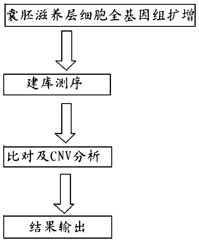
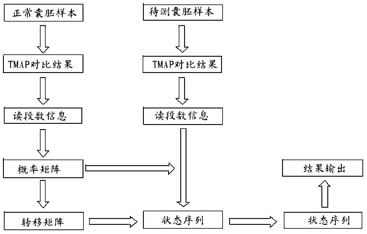
The invention relates to a method for detecting chromosome microdeletion and micro-duplication of a human embryo. The method comprises the following steps of performing whole genome amplification on cells cultured in vitro, interrupting DNA (deoxyribonucleic acid) molecules, and sequencing DNA fragments to obtain sequencing reads; comparing the sequencing reads with a reference sequence, and positioning the sequencing reads on the reference sequence; screening non-repeated areas of the reference sequence, and reserving the non-repeated areas; establishing a matrix of read number in windows through normal samples, analyzing the data of the normal samples, performing statistics on the read number of all the windows in the non-repeated areas, and establishing a probability matrix of the read number and chromosome enpeoids; calculating the copy number, i.e., the A / B / C state, of loci; selecting m continuous loci, i.e., the A state, as micro-duplication loci, and selecting m continuous loci, i.e., the C state, as microdeletion loci; contrasting the micro-duplication loci and the microdeletion loci with the existing CNV (copy number variation) and disease database, performing basic gene annotation and gene function analysis which relates to deletion parts, and annotating with a microdeletion syndrome disease type.
Description
Technical field [0001] The invention relates to the field of molecular cell biology, in particular to a method for detecting human embryo chromosome microdeletion and microduplication, which can detect the copy number variation of human embryo blastocyst trophoblast cell chromosome DNA fragments. Background technique [0002] Chromosome microdeletion / microduplication refers to a deletion or duplication with a length of 1.5kb-10Mb on a chromosome. Human chromosome microdeletion / microduplication syndrome is caused by the occurrence of small deletions or duplications in human chromosomes, that is, DNA fragment copy number variation, abbreviated as CNV, which causes complex phenotypic diseases, which can lead to serious diseases and abnormalities, such as congenital Sexual heart disease, growth retardation, limb deformities, etc. Common microdeletion syndromes include 22q11 microdeletion syndrome, cat barking syndrome, Angelman syndrome, AZF deletion and so on. [0003] The 2009 "Res...
Claims
Application Information
Patent Timeline
Application Date:The date an application was filed.
Publication Date:The date a patent or application was officially published.
First Publication Date:The earliest publication date of a patent with the same application number.
Issue Date:Publication date of the patent grant document.
PCT Entry Date:The Entry date of PCT National Phase.
Estimated Expiry Date:The statutory expiry date of a patent right according to the Patent Law, and it is the longest term of protection that the patent right can achieve without the termination of the patent right due to other reasons(Term extension factor has been taken into account ).
Invalid Date:Actual expiry date is based on effective date or publication date of legal transaction data of invalid patent.

