
Image processing device, image processing method, and image processing program

An image processing device and image technology, applied in image data processing, image enhancement, image analysis, etc., can solve the problems of low visual recognition and difficult observation of blood vessels

Active Publication Date: 2015-11-04
OLYMPUS CORP
View PDF10 Cites 5 Cited by
  • Summary
  • Abstract
  • Description
  • Claims
  • Application Information

AI Technical Summary

Problems solved by technology

[0003] However, in the case of observation using white light, there is a problem that the visibility of blood vessels existing in the superficial or deep layer of the mucous membrane is low, making it difficult to clearly observe

Method used


Image

  • Image processing device, image processing method, and image processing program
  • Image processing device, image processing method, and image processing program
  • Image processing device, image processing method, and image processing program

Examples


Embodiment approach 1

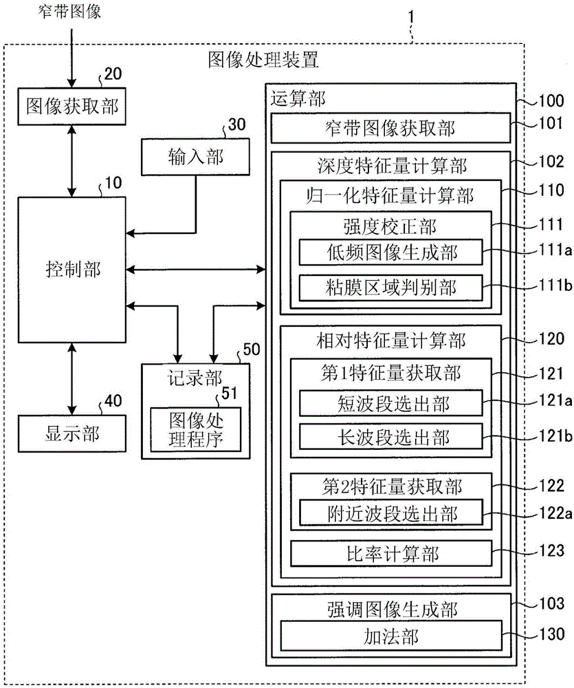
[0030] figure 1 It is a block diagram showing the image processing device according to Embodiment 1 of the present invention. The image processing device 1 according to Embodiment 1 is a device that performs image processing for estimating the depth of a blood vessel reflected in an image using at least three narrow-band images having different center wavelengths, and generating Intraluminal image with different tones to emphasize blood vessels. In addition, in the following description, although the narrow-band image acquired by the endoscope or the capsule endoscope in the lumen of the living body is taken as the processing object, it is also possible to use images other than the endoscope or the capsule endoscope. The image acquired by the observation device is used as the processing object.

[0031] As an example of a method of acquiring a narrow-band image with an endoscope, there is a method using an LED emitting light having a plurality of narrow-band wavelength peaks...

Embodiment approach 2

[0103] Next, Embodiment 2 of the present invention will be described.

[0104] Figure 10 It is a block diagram showing the configuration of the image processing device according to Embodiment 2 of the present invention. Such as Figure 10 As shown, the image processing device 2 of Embodiment 2 replaces figure 1 The computing unit 100 shown has a computing unit 200 . The configuration and operation of each unit of the image processing device 2 other than the computing unit 200 are the same as those in the first embodiment.

[0105] The calculation unit 200 includes a narrowband image acquisition unit 101 , a depth feature value calculation unit 202 , and an enhanced image generation unit 203 . However, the operation of the narrowband image acquisition unit 101 is the same as that of the first embodiment.

[0106] The depth feature calculation unit 202 has a normalized feature calculation unit 210 and a relative feature calculation unit 220 , and the depth feature calculat...


PUM


Abstract

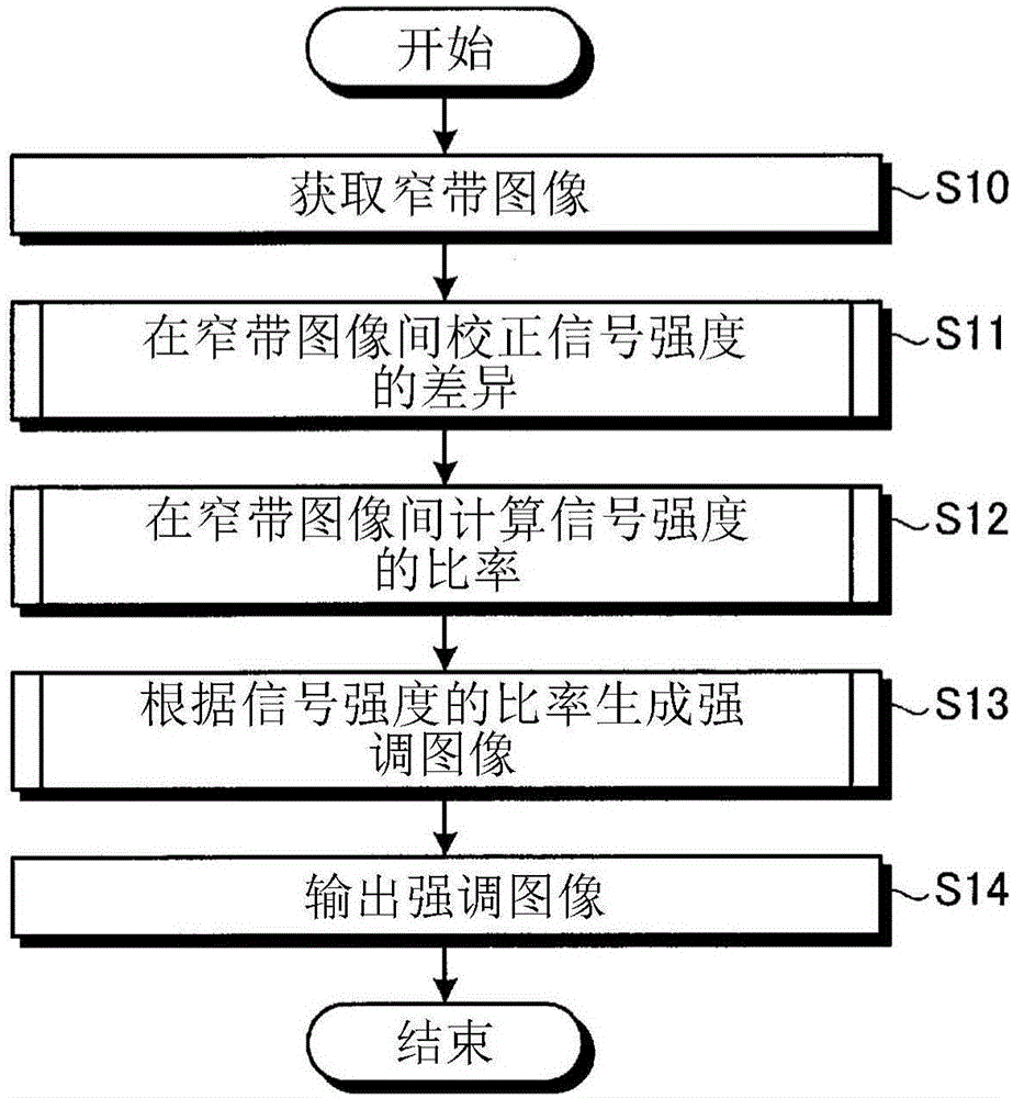
Provided are an image processing device, etc., whereby a blood vessel at a specific depth can be precisely extracted and enhanced from an image acquired by an endoscope. An image processing device (1) for processing an image acquired by imaging a living body, wherein the image processing device is provided with a narrow-band image acquiring unit (101) for acquiring at least three narrow-band images having mutually different center wavelengths, a depth characteristic amount calculation unit (102) for calculating a depth characteristic amount which is a characteristic amount correlated with the depth of a blood vessel in the living body on the basis of a difference in variation of a signal intensity due to variation in absorption of light irradiated to the living body in mutually different narrow-band images, and an enhanced-image creation unit (103) for creating an image in which the blood vessel is enhanced in accordance with the depth of the blood vessel on the basis of the depth characteristic amount.

Description

technical field [0001] The present invention relates to an image processing device, an image processing method, and an image processing program for image processing an image acquired by an endoscope that observes inside a lumen of a living body. Background technique [0002] In recent years, endoscopes have been widely used as medical observation devices capable of non-invasively observing the inside of a living body's lumen. Generally, a white light source such as a Xenon lamp is used as a light source of an endoscope, and the white light emitted from the light source is narrowed by combining a rotary filter with the light source to irradiate the inside of the lumen. The mirror is provided with a red filter, a green filter, and a blue filter that transmit light having wavelength bands of red light (R), green light (G), and blue light (B), respectively. The general shape and state of the mucous membrane in the lumen, the presence or absence of polyps, and the like can be ob...

Claims


Application Information

Patent Timeline
Patent Type & Authority Applications(China)
IPC IPC(8): A61B1/04A61B1/00A61B1/06H04N7/18
CPCA61B1/00186A61B1/00009G06T5/003G06T2207/10028G06T2207/10068G06T2207/30101H04N7/18A61B1/000094A61B1/044G06T5/73A61B1/041G06T7/0012G06T2207/10016G06T2207/10024G06T2207/20221
Inventor 弘田昌士神田大和
Owner OLYMPUS CORP
Learn More