Importing method of DICOM (Digital Imaging and Communications in Medicine) image

A technology of images and image files, applied in special data processing applications, instruments, electrical digital data processing, etc., can solve problems such as inability to quickly find image data

Active Publication Date: 2015-11-11
QINGDAO HISENSE MEDICAL EQUIP
View PDF5 Cites 22 Cited by
  • Summary
  • Abstract
  • Description
  • Claims
  • Application Information

AI Technical Summary

Problems solved by technology

[0005] Embodiments of the present invention provide a method for importing DICOM images to at least solve the problem in the prior art that the required image data cannot be quickly found during three-dimensional reconstruction of medical images

Method used


Image

  • Importing method of DICOM (Digital Imaging and Communications in Medicine) image
  • Importing method of DICOM (Digital Imaging and Communications in Medicine) image
  • Importing method of DICOM (Digital Imaging and Communications in Medicine) image

Examples


Embodiment 1

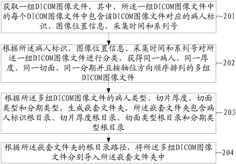
[0029] The embodiment of the present invention provides a method for importing a DICOM image, such as figure 2 shown, including:

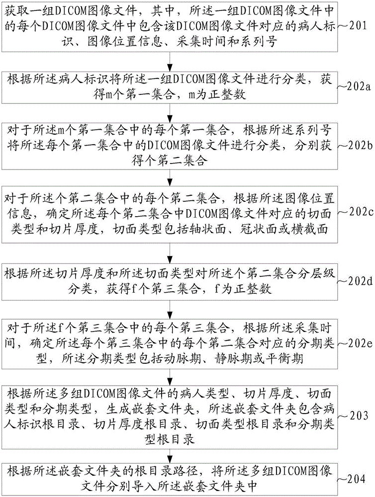
[0030] S201. Acquire a group of DICOM image files, wherein each DICOM image file in the group of DICOM image files includes the patient identification, image location information, acquisition time and serial number corresponding to the DICOM image file.

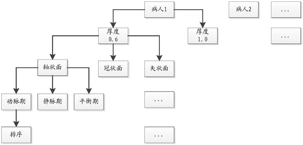
[0031] S202. Classify the set of DICOM image files according to the patient identification, image location information, acquisition time and serial number, and obtain multiple images of the same patient, the same thickness, the same section, the same stage and arranged in the order of the axial direction. Group DICOM image files.

[0032] S203. Generate a nested folder according to the patient type, slice thickness, slice type and staging type of the multiple groups of DICOM image files, where the nested folder includes a patient identification root directory, a slice thickness root directory, a...

Embodiment 2

[0112] An embodiment of the present invention is a device 50 for introducing a DICOM image, such as Figure 5 As shown, the introduction device 50 includes an acquisition unit 501 , a classification unit 502 , a generation unit 503 and an introduction unit 504 .

[0113] The acquiring unit 501 is configured to acquire a group of DICOM image files, wherein each DICOM image file in the group of DICOM image files includes the patient identification, image position information, acquisition time and series corresponding to the DICOM image file. No.

[0114] The classification unit 502 is configured to classify the set of DICOM image files according to the patient identification, image position information, acquisition time and serial number, and obtain the same patient, the same thickness, the same section, the same stage, and the axis Groups of DICOM image files arranged in bit-wise order.

[0115] The generating unit 503 is configured to generate a nested folder according to th...


PUM


Abstract

The invention provides an importing method of a DICOM (Digital Imaging and Communications in Medicine) image to solve the problem in the prior art that required image data can not be quickly found during the three-dimensional reconstruction of a medical image. The method comprises the following steps: obtaining one group of DICOM image files, wherein each DICOM image file in one group of DICOM image files comprises a patient identifier, image position information, acquisition time and a serial number corresponding to the DICOM image file; according to the patient identifier, the image position information, the acquisition time and the serial number, classifying one group of DICOM image files, obtaining multiple groups of DICOM image files of the same patient, the same thickness, the same tangent plane and the same stage, wherein the multiple groups of DICOM image files are sequentially arranged according to an axial position direction; according to the patient types, the section thicknesses, the tangent plane types and the stage types of the multiple groups of DICOM image files, generating a nesting folder; and according to a root directory path of the nesting folder, independently importing the multiple groups of DICOM image files into the nesting folder. The importing method is suitable for the field of the three-dimensional reconstruction of images.

Description

technical field [0001] The invention relates to the field of image three-dimensional reconstruction, in particular to a method for importing medical digital imaging and communication (Digital Imaging and Communications in Medicine, DICOM for short) images. Background technique [0002] 3D reconstruction of medical images is a technique for converting a set of 2D DICOM image sequences into 3D images. [0003] 3D reconstruction software generally requires a set of DICOM images to be of the same patient, the same thickness, the same section, the same stage, and arranged in the order of the axial direction. For example, if figure 1 As shown, during the three-dimensional reconstruction of medical images, patient 1 may be required, the slice thickness is 0.6, the axial plane, and a group of DICOM images in the arterial phase, the group of DICOM images are arranged in order in the axial direction. However, since there is no unified import standard in the current DICOM standard, t...

Claims


Application Information

Patent Timeline
Patent Type & Authority Applications(China)
IPC IPC(8): G06F17/30
CPCG06F16/16G06F16/583
Inventor 陈宗喜
Owner QINGDAO HISENSE MEDICAL EQUIP
Why Eureka
Try Eureka