
Method, device and equipment for obtaining ct images

A CT image and medical equipment technology, applied in the field of CT image acquisition, can solve problems such as CT image artifacts, projection errors, unevenness, etc., and achieve the effect of avoiding projection errors and scattering effects

Active Publication Date: 2018-08-21
NEUSOFT MEDICAL SYST CO LTD
View PDF10 Cites 0 Cited by
  • Summary
  • Abstract
  • Description
  • Claims
  • Application Information

AI Technical Summary

Problems solved by technology

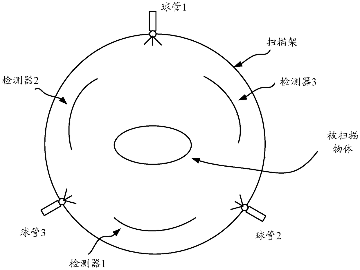
[0003] However, in the above method, when multiple tubes emit X-rays at the same time, the X-rays emitted by one tube will scatter when they irradiate the surface of the object, and the scattered rays will irradiate on the detector corresponding to the other tube. Projection errors are formed on the detector corresponding to the other tube, resulting in artifacts or inhomogeneities in the CT image

Method used


Image

  • Method, device and equipment for obtaining ct images
  • Method, device and equipment for obtaining ct images
  • Method, device and equipment for obtaining ct images

Examples


Embodiment Construction

[0045] The terminology used in this application is for the purpose of describing specific embodiments only, not to limit the application. As used in this application and the claims, the singular forms "a", "the" and "the" are intended to include the plural forms as well, unless the context clearly dictates otherwise. It should also be understood that the term "and / or" as used herein is meant to include any and all possible combinations of one or more of the associated listed items.

[0046] It should be understood that although the terms first, second, third, etc. may be used in this application to describe various information, the information should not be limited to these terms. These terms are only used to distinguish information of the same type from one another. For example, without departing from the scope of the present application, first information may also be called second information, and similarly, second information may also be called first information. Dependin...


PUM


Abstract

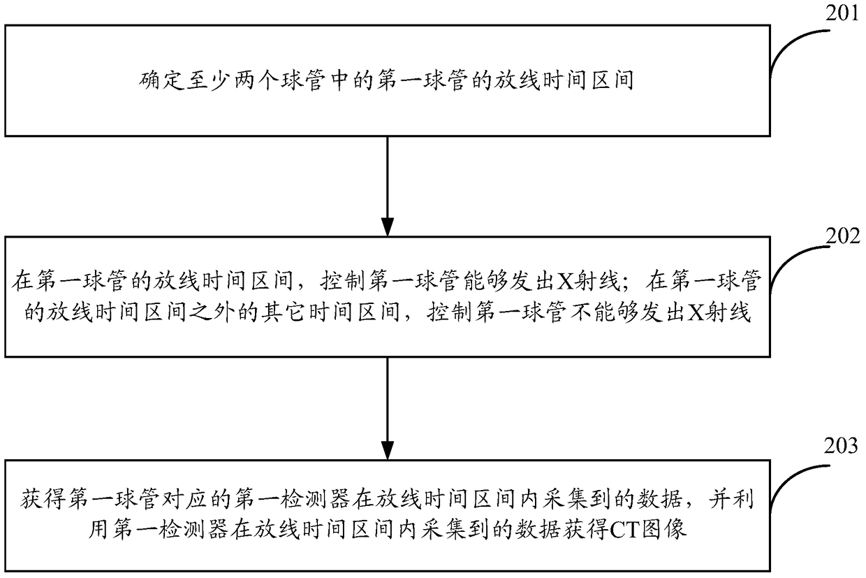
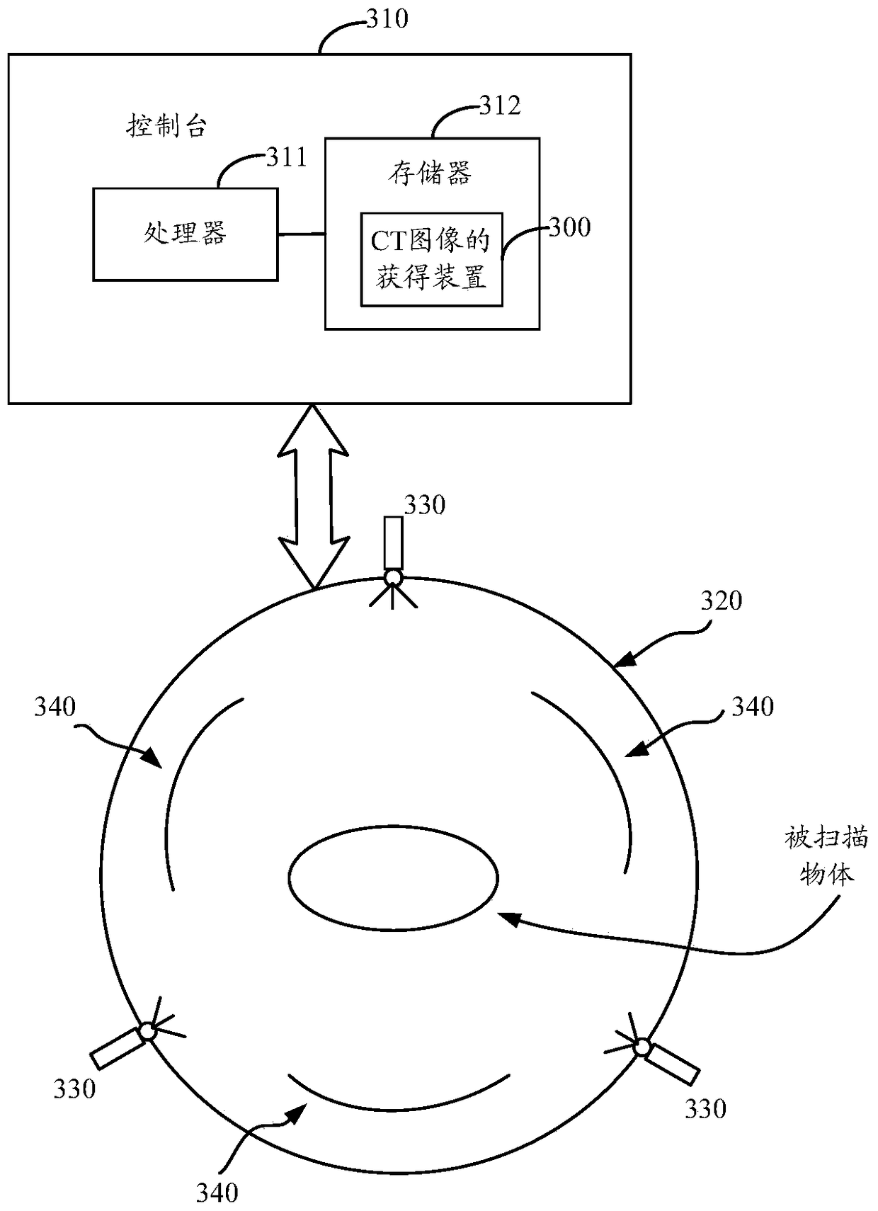
The invention discloses a CT image acquiring method, a CT image acquiring device and CT image acquiring equipment. The CT image acquiring method is applied on medical equipment. The medical equipment comprises at least two bulb tubes and at least two detectors which correspond with at least two bulb tubes. The CT image acquiring method comprises the steps of determining a ray emitting time interval of a first bulb tube in the two bulb tubes; in the ray emitting time interval, making the first bulb tube emit an X ray; in other time intervals except for the ray emitting time interval, preventing the first bulb tube to emit the X ray; acquiring data which are acquired by the a first detector which corresponds with the first bulb tube in the ray emitting time interval, and acquiring the CT image by means of the data which are acquired by the first detector in the ray emitting time interval. Through the CT image acquiring method, the CT image acquiring device and the CT image acquiring equipment, problems of irradiation of the emitted ray of one bulb tube to the detector which corresponds with the other bulb tube, projection error, etc. are prevented.

Description

technical field [0001] The present application relates to the technical field of medical equipment, in particular to methods, devices and equipment for obtaining CT images. Background technique [0002] For medical imaging medical equipment, such as CT (Computed Tomography, electronic computer tomography) equipment, in order to increase the scanning speed, multiple tubes and corresponding multiple detectors may be installed at different angles of the scanning frame , each tube corresponds to a detector. During the rotation of the scanning frame, multiple tubes will simultaneously emit X-rays, which will be received by the detectors corresponding to the tubes. Using this technology, the coverage angle range of X-ray projection can be doubled in the same time, so that the data required for CT image reconstruction can be obtained in a very short time. For moving objects (such as the heart, etc.) Imaging has great advantages. [0003] However, in the above method, when multip...

Claims


Application Information

Patent Timeline
Patent Type & Authority Patents(China)
IPC IPC(8): A61B6/03
Inventor 楼珊珊赵江魏
Owner NEUSOFT MEDICAL SYST CO LTD
Features
  • Generate Ideas
  • Intellectual Property
  • Life Sciences
  • Materials
  • Tech Scout
  • Unparalleled Data Quality
  • Higher Quality Content
  • 60% Fewer Hallucinations
Learn More