Method for detecting embryo chromosome abnormality by utilizing blastula culture solution

A chromosomal abnormality and culture medium technology, applied in the fields of biomedicine and molecular cell biology, can solve the problems of lifelong health to be observed, faulty test results, termination of embryonic development, etc., to avoid cell loss and damage, and simplify the operation.

Active Publication Date: 2016-03-02
XUKANG MEDICAL SCI & TECH (SUZHOU) CO LTD
View PDF4 Cites 32 Cited by
  • Summary
  • Abstract
  • Description
  • Claims
  • Application Information

AI Technical Summary

Problems solved by technology

[0008] 1. Cell sampling requires high technical requirements for embryo manipulation. If the operation is wrong or rough, it will cause severe damage to the embryo. If the damage is too serious, it will cause the termination of embryonic development.
[0009] 2. Even with good operation, cell loss and slight damage to embryos is inevitable when cell sampling
Although there is no evidence that cell loss and minor damage will have adverse effects on embryonic development and postnatal health, the technology is still young (only a few years), and whether it has long-term effects on human lifelong health remains to be seen. Observed
[0010] 3. In a few cases, the chromosomal status of several cells obtained by sampling is different from that of other cells in the embryo, resulting in incorrect test results

Method used


Image

  • Method for detecting embryo chromosome abnormality by utilizing blastula culture solution
  • Method for detecting embryo chromosome abnormality by utilizing blastula culture solution
  • Method for detecting embryo chromosome abnormality by utilizing blastula culture solution

Examples


Embodiment 1

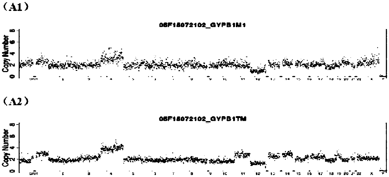
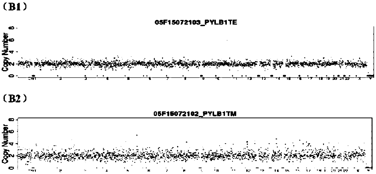
[0052] Select two in vitro fertilized embryo samples, A and B, and use blastocyst cell detection method and blastocyst culture fluid detection method to evaluate their chromosomal status respectively. The specific steps are as follows:

[0053] 1. Acquisition of Blastocyst Culture Medium

[0054] 1) After the fertilized eggs obtained by single sperm injection are cultured to the blastomere stage on the third day, the embryos are transferred to the newly prepared blastocyst culture microdrops for blastocyst culture.

[0055] 2) Aspirate the blastocyst-forming embryos and transfer them to a new blastocyst culture medium / or enter the vitrification process. The remaining original blastocyst culture medium (about 30ul) is sample A and sample B that need to be collected for PGS .

[0056] 2. Collection of blastocyst culture medium

[0057] 1) Place the collection tube containing 10 microliters of lysate (Tris-Cl40mM with a pH of 7.2, EDTA1mM, KCl15mM and 3% TritonX-100) at room te...


PUM


Abstract

The invention relates to a method for detecting embryo chromosome abnormality by utilizing blastula culture solution. By detecting free DNAs of an embryo source through embryo early stage in vitro solution, namely the blastula culture solution, trace DNAs undergo uniform whole genome amplification, a method of next generation sequencing and the like are utilized to analyze amplified DNA products, so that the chromosome condition of embryos, namely whether integral or local aneuploid of the chromosome arises, is judged. The blastula culture solution, which is waste at the embryo in vitro culture stage in the in vitro fertilization operation process, serves as a detection sample. According to the non-invasive method, cell damage to the embryos during conventional blastomere biopsy or blastula trophoblast cell biopsy sampling is avoided, no additional trouble is caused clinically, the operation during sample obtaining is simplified, and the safe reliability and simplicity are higher.

Description

technical field [0001] The invention relates to the fields of biomedicine and molecular cell biology, in particular to a method for detecting and analyzing the state of embryonic chromosomes using blastocyst culture fluid. Background technique [0002] Test-tube baby technology is a powerful technical means to combat infertility. The technical process is to first obtain multiple eggs (usually 8-15) from the mother, and then use the father's sperm to fertilize the eggs in vitro. When grown in in vitro culture medium for 5 days, the embryo is a cystic structure consisting of about 80 to 100 cells, namely the blastocyst. After placing 2-3 blastocysts into the mother's uterus, ideally, the blastocysts placed in the uterus can One to three successfully developed according to normal pregnancy until birth. However, due to various reasons, the success rate from blastocyst insertion into the uterus to fetal birth is not high, usually only about 40%. In addition to the mother's own h...

Claims


Application Information

Patent Timeline
Patent Type & Authority Applications(China)
IPC IPC(8): C12Q1/68G16B20/10G16B20/20G16B30/00
CPCC12Q1/6804C12Q1/6837C12Q1/6869G16B20/20G16B20/10G16B30/00Y02A90/10C12Q1/6809C12Q1/6853C12Q1/6806C12Q1/6883C12Q2521/537C12Q2525/161C12Q2527/101C12Q2527/125C12Q2531/113C12Q1/68C12Q1/686C12Q2525/204C12Q2521/531G16B20/00G16H50/20C12Q1/6811C12Q1/6827
Inventor 陆思嘉蔡立义姚兵
Owner XUKANG MEDICAL SCI & TECH (SUZHOU) CO LTD
Why Eureka
Try Eureka