Method for isolating tumor cell-derived exosomes from urine

A tumor cell and exosome technology, applied in the biological field, can solve the problems of difficult clinical application promotion, unsatisfactory, cumbersome operation, etc., and achieve the effects of large clinical application value, easy sampling and non-invasive, and easy to obtain in large quantities.

Active Publication Date: 2018-03-27
HANGZHOU LC BIOTECH
View PDF4 Cites 0 Cited by
  • Summary
  • Abstract
  • Description
  • Claims
  • Application Information

AI Technical Summary

Problems solved by technology

However, in fact, due to the very low content of exosomes in unit volume of urine, the exosomes obtained by a single extraction by the general method cannot meet the requirements of subsequent detection and sequencing. Secretion extraction method
In the few existing literatures related to the extraction of exosomes in urine, there are obviously many problems that make it impossible for clinical application
For example: The article entitled "Isolation and Purification of Exosomes in Urine" (Gonzales PA et al., Methods Mol Biol. 2010; 641:89-99) discloses two methods for isolating exosomes from urine, using ultrafast Exosomes are extracted by centrifugation or ultrafiltration tubes. Among them, the method of ultracentrifugation has more impurities and is very cumbersome to operate. It also needs to be equipped with an ultra-high-speed centrifuge, which is difficult to promote in clinical application; and the method of ultrafiltration tubes , although the use of ultra-high-speed centrifuges can be avoided, but the residues of other protein aggregation or macromolecular protein complexes cannot be avoided, and subsequent experimental research on exosomes will also bring wrong information
In addition, the origin of exosomes in urine is very complex, and it is impossible to assess the exosomes secreted by tumor cells
Based on the above limitations, there is currently no literature that has publicly reported the method of isolating tumor cell-derived exosomes (exosomes) from urine.

Method used


Image

  • Method for isolating tumor cell-derived exosomes from urine
  • Method for isolating tumor cell-derived exosomes from urine
  • Method for isolating tumor cell-derived exosomes from urine

Examples


Embodiment 1

[0052] Example 1: Extraction of exosome derived from tumor cells in the urine of patients with renal clear cell carcinoma

[0053] ①With the patient’s informed and ethical consent, collect 25ml of preoperative urine specimens from tumor patients (pathological biopsy has confirmed renal clear cell carcinoma) in a 50ml centrifuge tube, and centrifuge at 4°C and 300g for 10 minutes , separated to remove the cells in the urine, and then get the supernatant.

[0054] ② Freeze-dry the upper liquid obtained in ① to obtain a precipitate. Wherein, freeze-drying is completed in a freeze-drying apparatus, specifically: after freezing the upper layer liquid obtained in ① at -80° C., freeze-drying overnight (6 to 16 hours) on a freeze-drying apparatus at -40 to -50° C.

[0055] ③ Resuspend the lyophilized precipitate with 1ml of 1×PBS to obtain the first resuspension.

[0056] ④ Centrifuge the first resuspension at 4°C and 10,000 g for 30 minutes, separate to remove organelles and other ...

Embodiment 2

[0086] Example 2: Extraction of tumor-derived exosomes in the urine of patients with esophageal cancer

[0087] The tumor-derived exosome in the urine of patients with esophageal cancer was extracted by basically the same method as in Example 1. That is: ① The urine sample in ① is a 50ml urine sample from a patient with esophageal cancer before operation, and other steps are exactly the same as in Example 1.

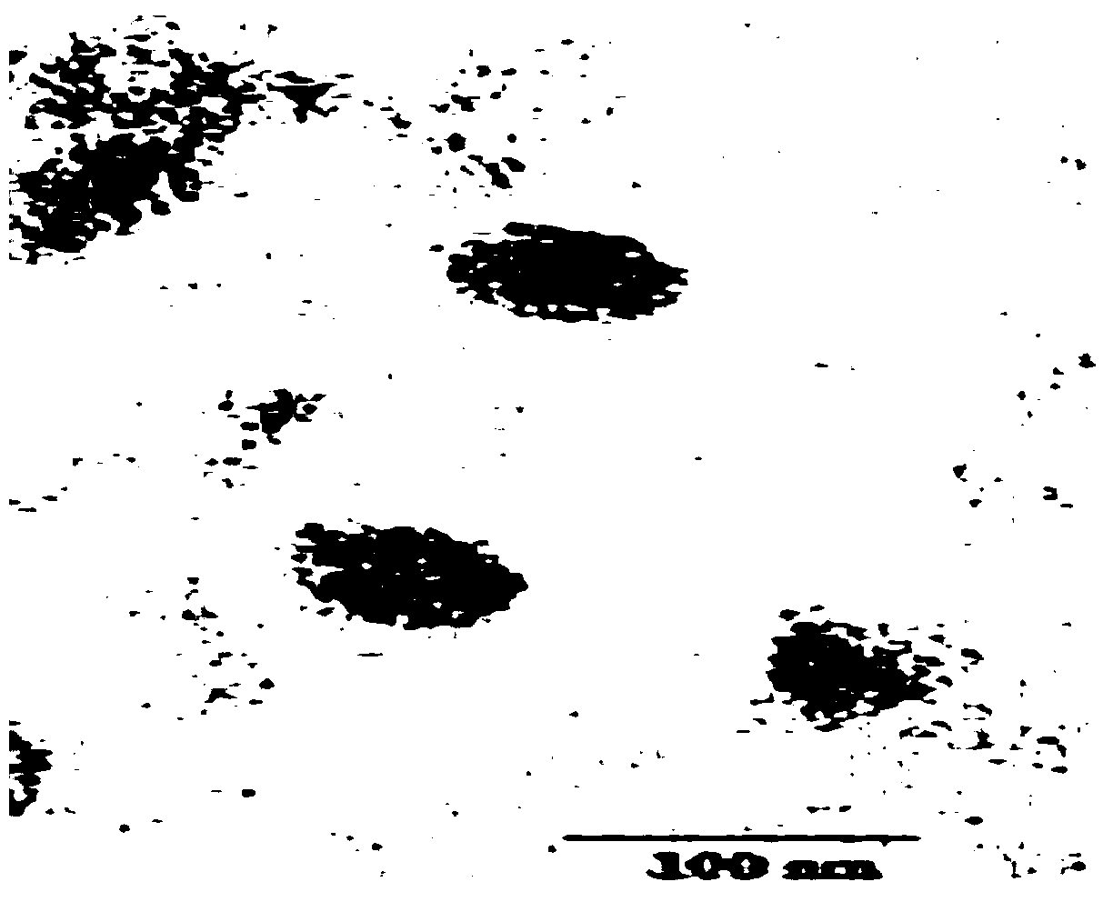
[0088] Similarly, the method in Example 1 was used to observe the product obtained in Example 2 with an electron microscope, and the morphology of exosomes consistent with the literature was also observed.

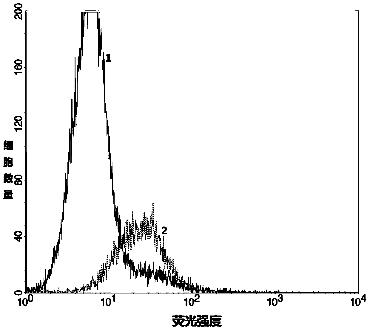
[0089] Further, the expression of CD63 and CD9 was detected by flow cytometry:

[0090] Resuspend the product obtained in Example 2 (i.e., magnetic bead-bound tumor cell-derived exosome) with 1 ml of 1×PBS to obtain 1 ml of esophageal cancer patient urine tumor-derived exosome resuspension, and resuspend the Add 5ul of flow cytometry antibodies CD63 and CD9 to the ...

Embodiment 3

[0093] Example 3: Extraction of tumor-derived exosomes in the urine of lung cancer patients

[0094] The tumor-derived exosome in 25 ml of urine of a patient with lung cancer was extracted by basically the same method as in Example 1. That is: ① The urine sample in the middle is the urine sample of the patient with lung cancer before operation, and other steps are exactly the same as in Example 1.

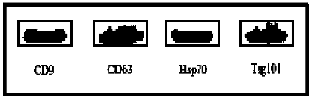
[0095] Using the product characterization method in Example 1, the product obtained in Example 3 was observed and tested. The results showed that: the product obtained in Example 3 had an exosome morphology that was consistent with that recorded in the literature, and the unique characteristics of exosomes were detected in the product. The expression of proteins CD9, CD63, Hsp70, Tsg101 and the strong expression of tumor antigen EpCAM protein prove that the product obtained in Example 3 is the exosome derived from tumor cells extracted from urine.


PUM

PropertyMeasurementUnit
volumeaaaaaaaaaa

Abstract

The invention discloses a method for separating tumor cell derived-exosomes from urine. The method comprises the following steps: freeze-drying a urine sample, extracting total exosomes by using an extracting reagent, adsorbing by using EpCAM antibody labeled magnetic beads, and separating and purifying the tumor cell derived-exosomes. The method overcomes the defect of extraction of a small amount of urine and unable reaching of large scale screening or expressing detection experiment requirements of present extraction kits, realizes obtaining of concentration-detectable exosomes in urine, and also overcomes the defect of low exosome tumor specificity of present detection in order to accurately detect the bio-information of the tumor cell derived-exosomes in the urine. The method has the advantages of noninvasive sampling, strong specificity, convenience, easy implementation and low price, can be directly used in subsequent detection, and has high clinic popularization application values.

Description

technical field [0001] The invention belongs to the field of biotechnology, and in particular relates to a method for isolating tumor cell-derived exosomes (exosomes) from urine. Background technique [0002] Exosomes are nanoscale vesicles that shuttle between cells with a bilayer. Exosomes can be secreted by a variety of cells, and exosomes secreted by different cells can have similar or different characteristics and biological functions. Tumor cells can also release exosomes, and during the growth process of tumors, exosomes will continue to be released outside the cells to affect the tumor microenvironment, and their role in tumorigenesis and development has attracted more and more attention. Exosomes can not only exist stably in body fluids such as serum, plasma, and urine, but also selectively wrap / release genetic information (miRNA, protein, etc.) in their cells, so extracellular exosomes can be extracted for use in diseases The diagnosis and monitoring of tumors an...

Claims


Application Information

Patent Timeline
Patent Type & Authority Patents(China)
IPC IPC(8): C12N5/09
CPCG01N1/28
Inventor 赵安凌志强毛伟敏
Owner HANGZHOU LC BIOTECH
Why Eureka
Try Eureka