Method for medical ultrasound three-dimensional imaging based on parallel computer

A 3D imaging and computer technology, applied in the field of medical ultrasound image processing and image display, can solve the problems of large amount of data, increasing the complexity of 3D reconstruction algorithms, and the spatial positions of 2D image sequences are not regularly arranged, etc.

Inactive Publication Date: 2012-12-26
SOUTH CHINA UNIV OF TECH
View PDF3 Cites 11 Cited by
  • Summary
  • Abstract
  • Description
  • Claims
  • Application Information

AI Technical Summary

Problems solved by technology

The reconstruction algorithm of this method is simple and fast, but the scanning method is limited by a lot of rules, which makes its application range relatively narrow
Free-arm scanning can scan in any position and direction, which provides greater flexibility to the operator. However, the spatial positions of the two-dimensional image sequences obtained by this scanning method are not regularly arranged, and the amount of data may be large, which not only increases the Increasing the complexity of the 3D reconstruction algorithm will also increase the time of the entire reconstruction process, which is a relatively big problem for ultrasound 3D imaging technology that requires high clinical real-time performance

Method used


Image

  • Method for medical ultrasound three-dimensional imaging based on parallel computer
  • Method for medical ultrasound three-dimensional imaging based on parallel computer
  • Method for medical ultrasound three-dimensional imaging based on parallel computer

Examples


Embodiment

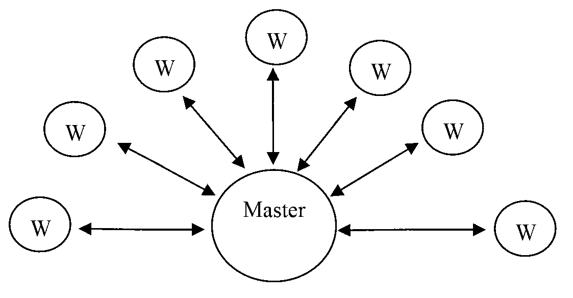
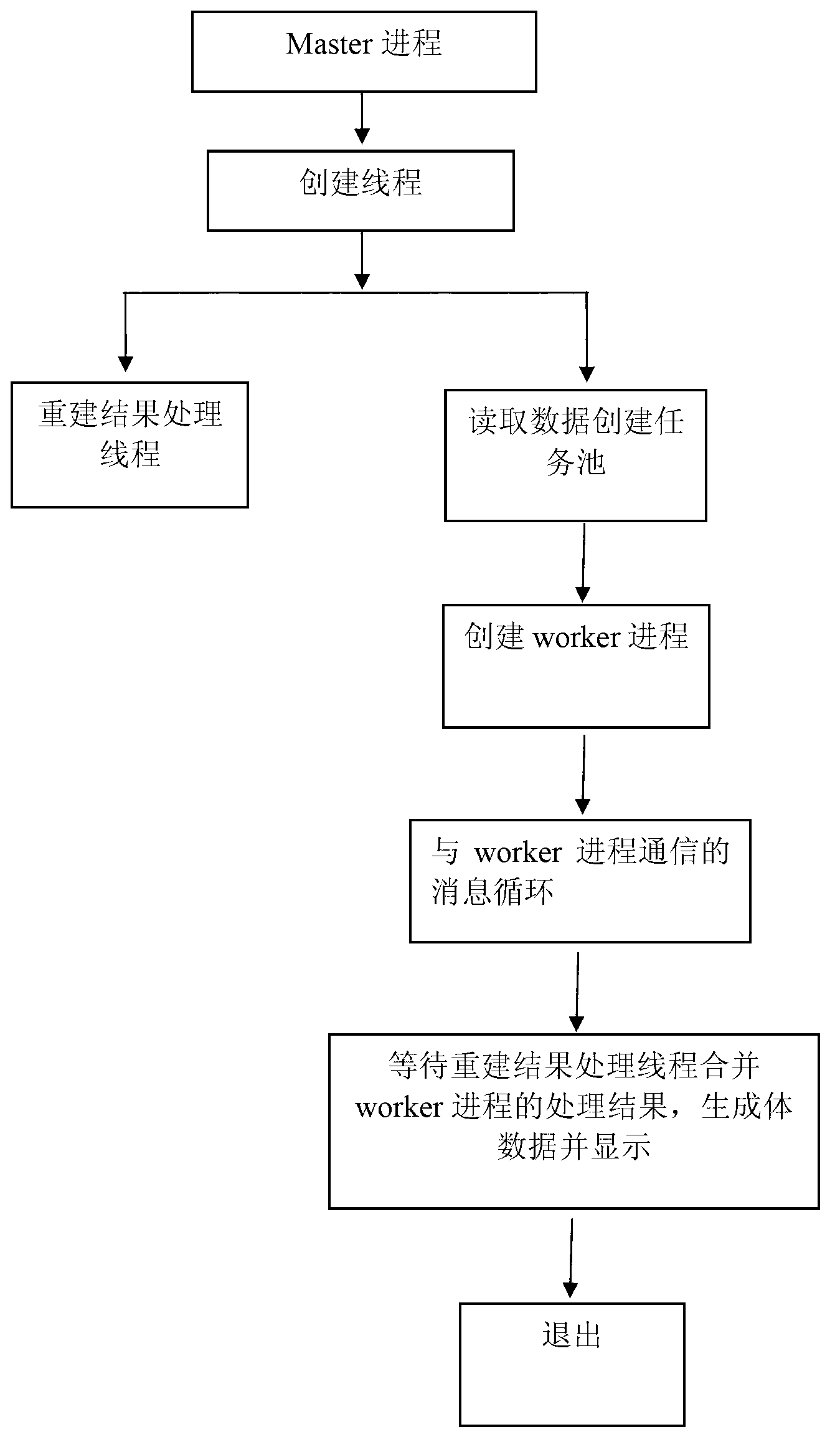
[0048] This implementation requires the AVI ultrasonic image video files collected by the ultrasonic equipment, use the MPI-based computer cluster as a parallel computing platform for three-dimensional imaging, use a flat-screen display to display the user graphical interface, and use C and C++ languages ​​to compile various processing programs , the present invention can be better implemented.

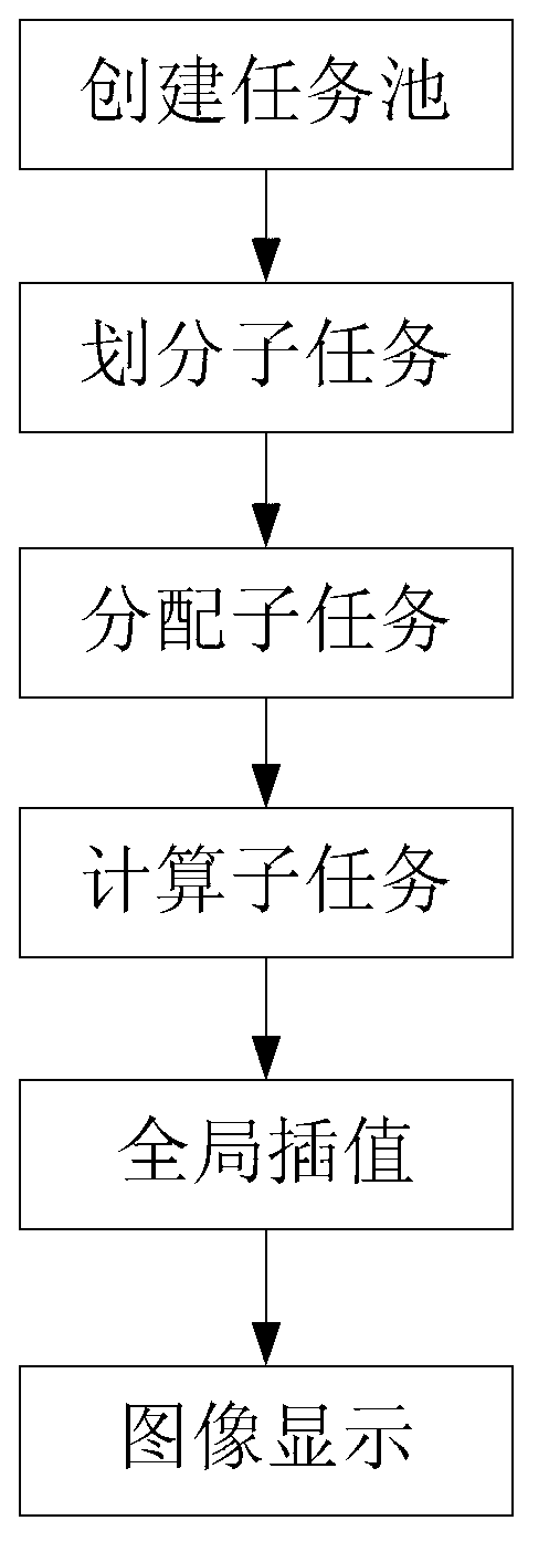
[0049] Such as figure 1 As described, the present embodiment is a parallel computer-based medical ultrasound three-dimensional imaging method, comprising the following steps:

[0050] S1. Create a task pool, read image data and build a task pool according to user-set parameters;

[0051] S2, dividing the subtasks, adopting the method of domain division to divide the ultrasonic image sequence collected into a plurality of subtasks according to the defined granularity, each subtask is to complete the interpolation of adjacent images of S frames;

[0052] S3, assigning subtasks, and as...


PUM


Abstract

The invention discloses a method for medical ultrasound three-dimensional imaging based on parallel computers. The method includes the following steps: S1. creating a task pool, namely, reading image data in accordance with user setting parameters and construct the task pool; S2. conduct partitioning to obtain sub-tasks, namely, adopting domain partition method to partition collected ultrasound image sequences into a plurality of sub-tasks in accordance with defined particle size, wherein each sub-task is an interpolation to complete adjacent images of S frames; S3. sub-tasks distribution, namely, distributing sub-tasks to different processors; S4. calculating sub-tasks, namely each processor executing sub-tasks coordinately and parallelly; S5. overall interpolation, namely conducting overall interpolation to sub-task results obtained by reconstruction of each processor to get a three-dimensional image; and S6. image display, namely displaying the reconstructed three-dimensional image. The method solves the time-consuming problem of the ultrasound three-dimensional imaging. Besides, the parallel computer environment is constructed by low-cost computers. Thus, the computing resources can be fully used and the cost is also low.

Description

technical field [0001] The invention relates to the technical field of computer-aided medical imaging, in particular to a method for medical ultrasonic image processing and image display. Background technique [0002] Ultrasound imaging has been widely used in clinical practice due to its advantages of convenience, no trauma, no pain, no ionizing radiation and low cost. Among them, three-dimensional ultrasound imaging technology has been more and more used in ultrasound diagnosis because of its intuitive image display and high diagnostic accuracy. At present, the scanning methods commonly used in medical 3D ultrasound imaging include: mechanical positioning parallel scanning, sector scanning, rotating scanning and free-arm (Free-Hand) scanning. Among them, mechanical positioning parallel scanning, sector scanning and rotating scanning are to use the existing two-dimensional ultrasonic diagnostic equipment (probe) combined with some positioning machinery to obtain a series o...

Claims


Application Information

Patent Timeline
Patent Type & Authority Applications(China)
IPC IPC(8): A61B8/00G06T17/00
Inventor 黄庆华胡伟
Owner SOUTH CHINA UNIV OF TECH
Why Eureka
Try Eureka