Blood vessel extraction method

An extraction method and blood vessel technology, applied in the field of medical tomography image processing, can solve the problems of multi-scanning radiation dose and time-consuming of patients

Active Publication Date: 2016-03-23
SHANGHAI UNITED IMAGING HEALTHCARE
View PDF11 Cites 25 Cited by
  • Summary
  • Abstract
  • Description
  • Claims
  • Application Information

AI Technical Summary

Problems solved by technology

This method needs to scan the patient twice, which is time-co

Method used


Image

  • Blood vessel extraction method
  • Blood vessel extraction method
  • Blood vessel extraction method

Examples


Embodiment Construction

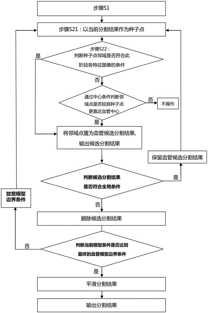
[0032] The present invention will be described in further detail below in conjunction with the accompanying drawings and specific embodiments. Advantages and features of the present invention will be apparent from the following description and claims. It should be noted that the drawings are all in a very simplified form and use imprecise ratios, which are only used to facilitate and clearly assist the purpose of illustrating the embodiments of the present invention.

[0033] In CT angiography (CTA), accurate vessel segmentation techniques are crucial for vessel analysis and disease diagnosis. The borders of blood vessels in the head and neck are mostly adhered to the bones, making segmentation and extraction difficult. Embodiments of the present invention aim at accurately segmenting and extracting blood vessels, especially head and neck blood vessels, from the original image. Based on the premise of having a center line, the present invention can quickly and accurately segm...


PUM


Abstract

The invention provides a blood vessel extraction method. The blood vessel extraction method comprises steps that S1, a blood vessel center line in an original blood vessel image is extracted, a to-be-segmented blood vessel area is acquired according to the center line, the center line is set as an initial segmentation result, and boundary values of the blood vessel at the original image and at least one characteristic image are initialized and taken as blood vessel model conditions; S2, the present segmentation result is taken as a seed point, whether original image values and at least one characteristic image value of neighborhood points adjacent to the seed point satisfy the present blood vessel model condition is determined, if yes, the neighborhood points are taken as candidate blood vessel segmentation results. Through the method, blood vessel extraction can be more accurately extracted.

Description

technical field [0001] The invention relates to the processing of medical tomography images, in particular to a blood vessel extraction method in angiography. Background technique [0002] In Computed Tomography Angiography (CTA for short), it is necessary to separate blood vessel images from images of other tissues to extract blood vessels for further observation and analysis of blood vessels. Existing techniques for vessel extraction in CTA are mainly divided into two categories: methods based on standard vessel models and methods not based on standard vessel models. [0003] US Patent Document 1: Jean-MichelRouet, FranckLaffargueandMaximFradkin, Imageprocessingsystemforautomaticsegmentationofa3-Dtree-liketubularsurfaceofanobject, using3-Ddeformablemeshmodels.USPatent, 2008, US2008 / 0094389A1 provides a variable grid model method, which needs to provide a standard vessel model first, and then calculate the center of the vessel Line, and then according to the center line, u...

Claims


Application Information

Patent Timeline
IPC IPC(8): G06T7/00
CPCG06T7/0012G06T2207/30101
Inventor 王策
Owner SHANGHAI UNITED IMAGING HEALTHCARE
Why Eureka
Try Eureka