

Lung segmentation method
A lung and regional technology, applied in the medical field, can solve the problems of large amount of calculation, difficulty in building models, long processing time, etc., and achieve the effect of improving the effect, preventing background adhesion, and speeding up the segmentation speed
- Summary
- Abstract
- Description
- Claims
- Application Information
AI Technical Summary
Problems solved by technology
Method used
Image

Examples
Embodiment Construction
[0030] The present invention will be described in further detail below in conjunction with the accompanying drawings and specific embodiments. Advantages and features of the present invention will be apparent from the following description and claims. It should be noted that the drawings are all in a very simplified form and use imprecise ratios, which are only used to facilitate and clearly assist the purpose of illustrating the embodiments of the present invention.
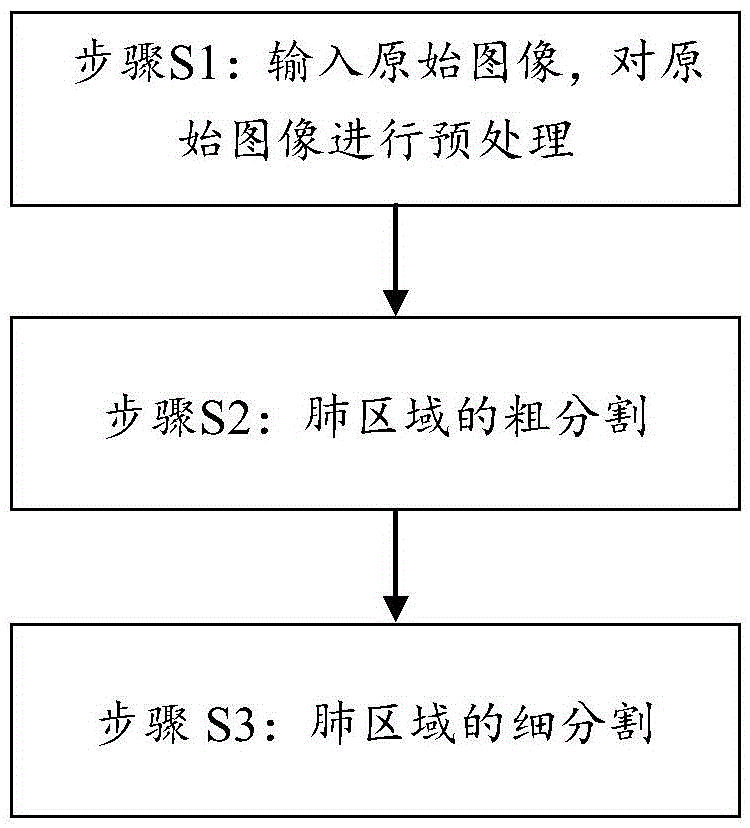
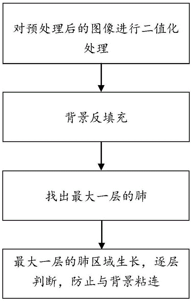
[0031] Please refer to figure 1 As shown, the lung segmentation method in the embodiment of the present invention includes the following steps:
[0032] Step S1: Input the original image, and preprocess the original image.
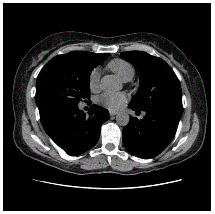
[0033] Such as image 3 As shown, the original image is a chest CT image obtained by scanning with CT equipment and conforming to the DICOM3.0 standard. The method used in the preprocessing is to perform noise reduction processing on the original image, such as Gaussian smoothing proces...
PUM
 Abstract
Description
Claims
Application Information