a protein chip
A protein chip, chip technology, applied in instruments, biological material analysis, measurement devices, etc., can solve the problems of undetectable technology, false negative results, difficulty in diagnosing cancer, etc., and achieve the effect of improving sensitivity and reducing preparation costs
- Summary
- Abstract
- Description
- Claims
- Application Information
AI Technical Summary
Problems solved by technology
Method used
Image

Examples
Embodiment 2
[0039] Preparation of various polymer plates with immunocompetent wells
[0040] 1. Prepare small hole arrays on various plates: use hot pressing or drilling to form holes, the diameter of the holes is 3-6mm small holes, the volume is preferably 10-50 microliters, and the hole distance depends on the measuring instrument. Suitable microtiter plates can also be used.
[0041] 2. Coating primary antibody: Add 10-50 microliters of appropriately diluted primary antibody to each well at pH 7.0-9.5, incubate overnight at 4°C or 2 hours at 37°C, and adsorb the primary antibody on the plate On the wall of the small hole, suck out the antibody solution after the reaction.
[0042] 3. Blocking: Add 10-50 microliters of blocking solution to each well, incubate at 37°C for 2 hours, or overnight at 4°C. After the reaction, the blocking solution was shaken off, air-dried and sealed at 4°C for future use.
[0043] Of course, the immunological microassay plate is used to prepare the polyme...
Embodiment 3
[0045] Commonly used polymer balls include polypropylene balls, nylon balls, polystyrene balls and their microspheres. The diameter of the high molecular polymer ball is between 4-6mm, and the diameter of the microsphere is between 10-60nm. Some of the balls have a smooth surface and some have a rough surface.
[0046] (1) Polypropylene beads or microspheres The primary antibody can be coated on the surface of the beads or microspheres by electrostatic adsorption, see Example 2 for details.
[0047] (2) React the nylon ball with 1-4 mol / L hydrochloric acid for 1-6 hours at room temperature to expose the amino and carboxyl groups, and then coat the primary antibody on the surface of the nylon ball with carbodiimide or glutaraldehyde .
[0048] (3) Polystyrene beads or microspheres coated with the first antibody The first antibody can be coated on the surface of polystyrene beads or microspheres by electrostatic adsorption, see Example 2; polystyrene beads Balls or microspher...
Embodiment 4
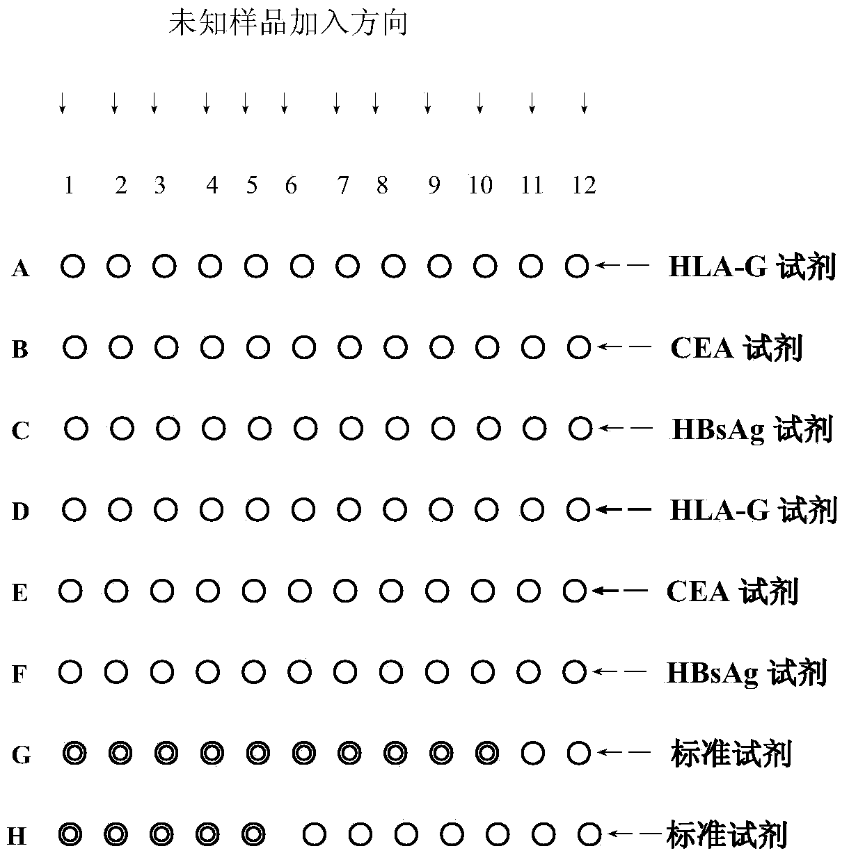
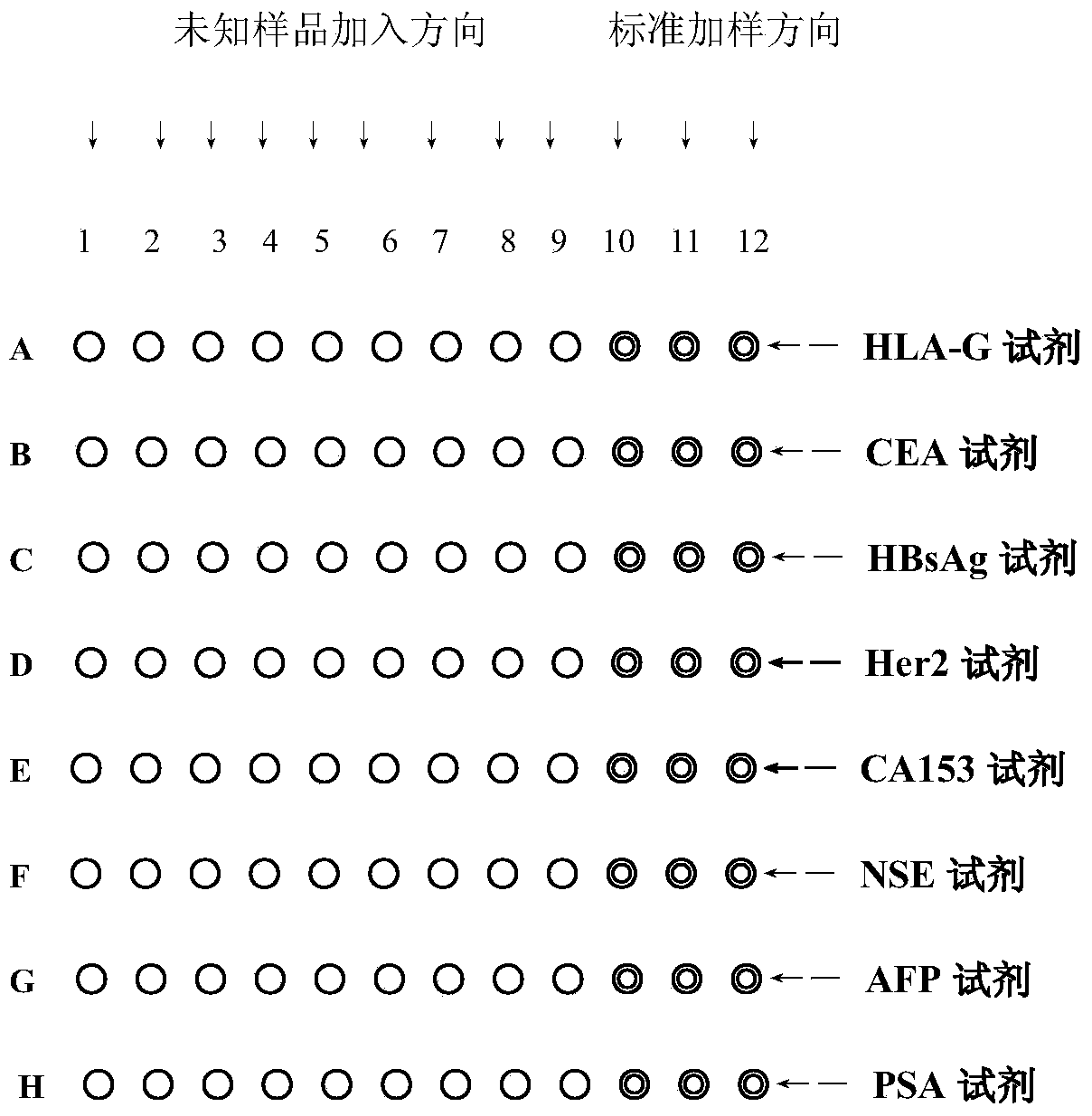
[0050] Using the glass protein chip and polymer chip prepared in embodiment 1 and embodiment 2 to measure HLA-G
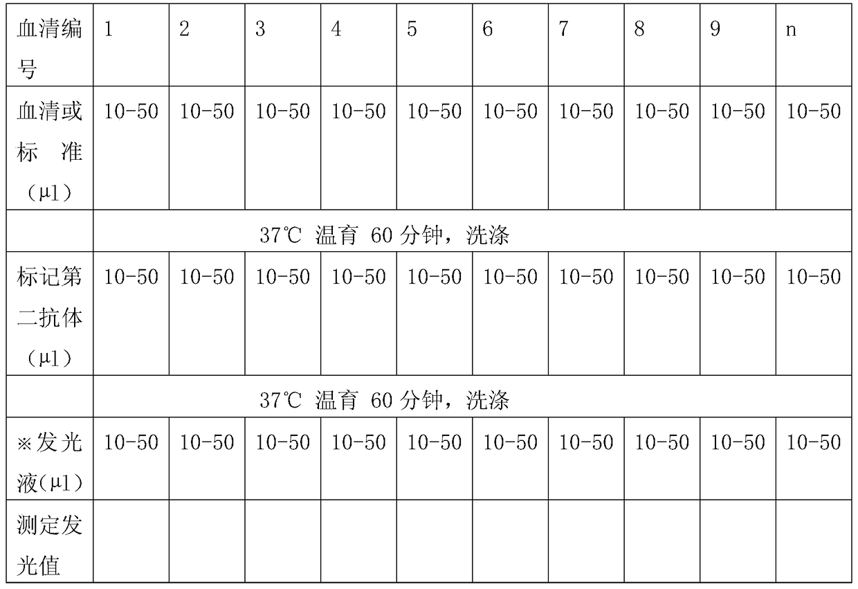
[0051] 1. Determination of the serum to be tested: add the No. 1 serum to be tested to each of the first vertical row of the glass slide (or ball) or the polymer plate after binding the first antibody and blocking the glass chip or polymer chip. In the active wells, serum No. 2 is added to each active well in the second vertical row, and so on, serum No. 3 to serum n are added to each active well in the third to nth vertical rows in turn, and each active well is added 10-50 microliters of serum to be tested.
[0052] 2. Incubate the active glass slide (or ball) or polymer plate (or ball) at 37°C for 60 minutes, shake once (5 seconds) in the middle of the reaction, suck out the serum, and discard it after disinfection.
[0053] 3. Washing: Add 10-50 microliters of washing solution to each active well, wash 5-8 times, shake for 5 seconds each time, suck out the wash...
PUM
| Property | Measurement | Unit |
|---|---|---|
| diameter | aaaaa | aaaaa |
| diameter | aaaaa | aaaaa |
Abstract
Description
Claims
Application Information
 - R&D Engineer
- R&D Manager
- IP Professional
- Industry Leading Data Capabilities
- Powerful AI technology
- Patent DNA Extraction
Browse by: Latest US Patents, China's latest patents, Technical Efficacy Thesaurus, Application Domain, Technology Topic.
© 2024 PatSnap. All rights reserved.Legal|Privacy policy|Modern Slavery Act Transparency Statement|Sitemap