Supercharge Your Innovation With Domain-Expert AI Agents!

Scanning data processing method and device

A technology of scanning data and processing methods, which is applied in the medical field and can solve problems such as inaccurate positioning

Active Publication Date: 2016-06-22
上海伴诚医疗科技有限公司
View PDF10 Cites 1 Cited by
  • Summary
  • Abstract
  • Description
  • Claims
  • Application Information

AI Technical Summary

Problems solved by technology

[0003] The object of the present invention is to provide a scan data processing method and device, which can process the scan data of the head to obtain a bracket model with a positioning channel for surgery on the head lesion, so that in the above-mentioned existing operation process The problem of inaccurate positioning due to manual measurement and positioning has been improved

Method used


Image

  • Scanning data processing method and device
  • Scanning data processing method and device
  • Scanning data processing method and device

Examples


no. 1 example

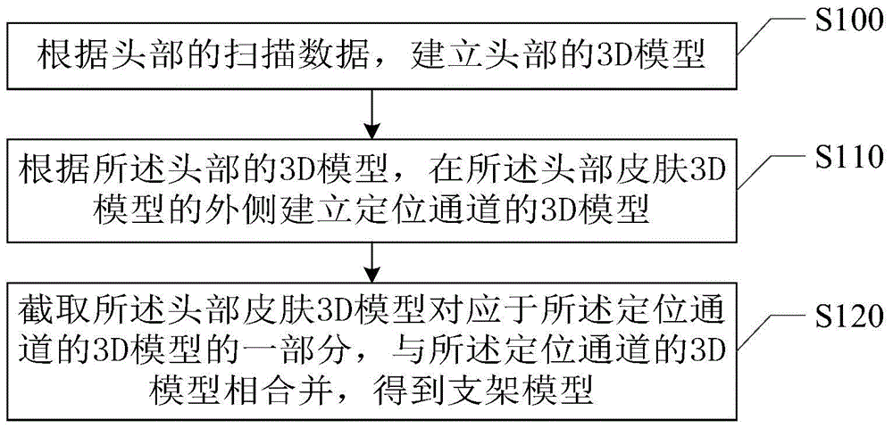
[0023] Such as figure 1 As shown, the embodiment of the present invention provides a scanning data processing method, the method comprising:

[0024] Step S100: Establish a 3D model of the head according to the scan data of the head, the 3D model of the head includes a 3D model of the head skin corresponding to the skin of the head and a 3D model of the head lesion corresponding to the head lesion;

[0025] Scan the head that needs to undergo related operations to obtain scan data. The file corresponding to the scan data is a dicom file, and the dicom file is synthesized into a 3D model, that is, converted into a file corresponding to the 3D model, and the 3D corresponding to the corresponding head is obtained. Model. The head 3D model includes a head lesion 3D model and a head skin 3D model. Among them, the 3D model of the head lesion includes a hematoma 3D model and a tumor 3D model.

[0026] In addition, scanning methods include CT scanning and MR scanning, and correspon...

no. 2 example

[0060] Such as Figure 6 As shown, the scan data processing method provided in this embodiment includes:

[0061] Step S200: Create a 3D model of the head according to the scan data of the head, the 3D model of the head includes a 3D model of the skin of the head corresponding to the skin of the head and a 3D model of the lesion of the head corresponding to the lesion of the head.

[0062] In this embodiment, the scan data of the head includes CT scan data, the 3D model of the head lesion includes a 3D model of hematoma, and the step of establishing the 3D model of the head according to the scan data of the head includes: The 3D model of the head synthesized from the CT scan data; wherein, the data part of the grayscale value from -65 to -104 in the CT scan data corresponds to the 3D model of the head skin, and the grayscale value in the CT scan data is The data portion with values ​​65 to 82 corresponds to the 3D model of the hematoma.

[0063] In addition, in this embodime...

no. 3 example

[0077] Such as Figure 7 As shown, this embodiment provides a scan data processing method device 300, the device includes:

[0078] The 3D model acquisition module 310 is configured to establish a 3D model of the head according to the scan data of the head, and the 3D model of the head includes a 3D model of the head skin corresponding to the head skin and a head skin corresponding to the head lesion. Lesion 3D model;

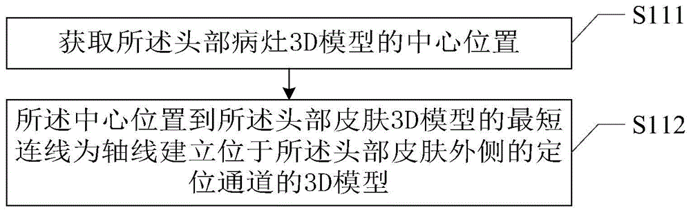
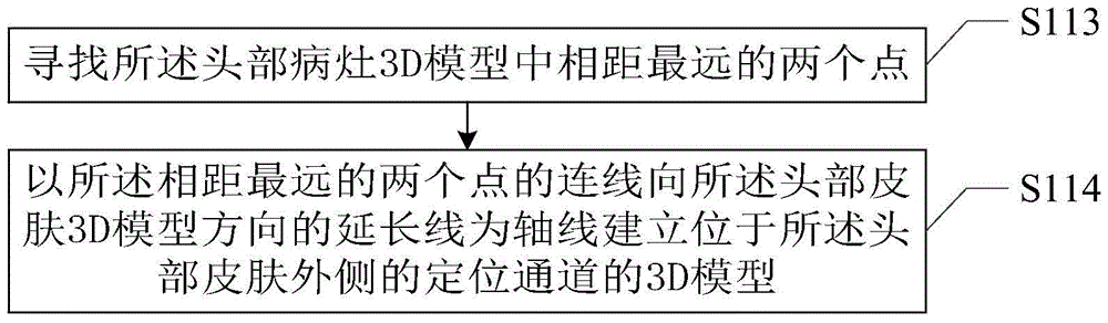
[0079] The positioning channel establishment module 320 is configured to establish a 3D model of the positioning channel on the outside of the 3D skin model of the head according to the 3D model of the head, and the axis of the 3D model of the positioning channel passes through the lesion of the head 3D model;

[0080] The bracket model acquisition module 330 is configured to intercept a part of the 3D model of the head skin corresponding to the positioning channel to obtain a useful 3D model of the head skin, and combine the useful 3D model of the head skin ...


PUM


Abstract

The invention relates to the field of medicine and provides a scanning data processing method and device. The method comprises the steps of building a 3D model of a head according to the scanning data of the head, wherein the 3D model of the head includes a head skin 3D model corresponding to the head skin and a head focus 3D model corresponding to a head focus; according to the 3D model of the head, building a locating channel 3D model on the outer side of the head skin 3D model, the axis of the locating channel 3D model passing through the head locus 3D model; cutting out the part, corresponding to the locating channel 3D model, of the head skin 3D model to obtain a useful head skin 3D model; combining the useful head skin 3D model and the locating channel 3D model to obtain a support model for the locus. The method is accurate in locus locating and is simple and convenient.

Description

technical field [0001] The present invention relates to the field of medicine, in particular to a scan data processing method and device. Background technique [0002] Cerebral hemorrhage and brain tumors are frequently-occurring diseases in our country, and they are also one of the important diseases that cause disability and death to our citizens. At present, the treatment of cerebral hemorrhage adopts surgery and minimally invasive channel technology. However, regardless of the means, the location of the hematoma before the operation is often manually measured and positioned, which often leads to errors in the location, thereby increasing the difficulty of the operation and operation, and even affecting the treatment effect, resulting in a poor treatment effect and bringing great harm to the patient and his family. It is a huge risk and brings a serious burden to the society. For brain tumors, primary hospitals are often less equipped with expensive navigation assistanc...

Claims


Application Information

Patent Timeline
Patent Type & Authority Applications(China)
IPC IPC(8): A61B5/00G06T17/00
CPCA61B5/72G06T17/00G06T2207/10081G06T2207/30096
Inventor 兰津刘奇汪宇跃
Owner 上海伴诚医疗科技有限公司
Features
  • R&D
  • Intellectual Property
  • Life Sciences
  • Materials
  • Tech Scout
  • Unparalleled Data Quality
  • Higher Quality Content
  • 60% Fewer Hallucinations
Learn More