A method and device for detecting immunogen

A detection method and immunogen technology, applied in the field of biomedicine, can solve problems such as severe cross-reactivity, poor specificity, and inability to distinguish HPV types

Active Publication Date: 2018-11-16
ATTOGEN BIOMEDICAL SUZHOU INC
View PDF11 Cites 1 Cited by
  • Summary
  • Abstract
  • Description
  • Claims
  • Application Information

AI Technical Summary

Problems solved by technology

In addition, various methods for detecting HPV in the prior art have severe cross-reactivity and poor specificity, and cannot distinguish specific HPV types

Method used


Image

  • A method and device for detecting immunogen
  • A method and device for detecting immunogen
  • A method and device for detecting immunogen

Examples


preparation example Construction

[0087] Preparation of monoclonal antibodies

[0088] Antibodies suitable for use in the present invention can be prepared by various techniques known to those skilled in the art. For example, an antigen of the invention may be administered to an animal to induce the production of monoclonal antibodies. For monoclonal antibodies, hybridoma technology can be used to prepare (see Kohler et al., Nature 256; 495, 1975; Kohler et al., Eur.J.Immunol.6:511, 1976; Kohler et al., Eur.J.Immunol. 6:292,1976; Hammerling et al., In Monoclonal Antibodies and T Cell Hybridomas, Elsevier, N.Y., 1981) or can be prepared by recombinant DNA methods (US Patent No. 4,816,567).

[0089] Representative myeloma cells are those that fuse efficiently, support stable high-level production of antibody by selected antibody-producing cells, and are sensitive to culture medium (HAT medium matrix), including myeloma cell lines, such as murine Myeloma cell lines, including those derived from MOPC-21 and MPC-...

Embodiment 1

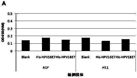
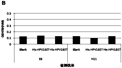
[0143] Example 1 Specific Identification of HPV18 E7 Protein

[0144] In this example, monoclonal antibodies H11 and F1 are taken as examples to illustrate the specific experimental methods in this example.

[0145]Detect the reaction of monoclonal antibodies H11 and F1 to His-HPV18 E7 and His-HPV16 E7 proteins, and use the multi-antibody coating sandwich ELISA method for identification:

[0146] (1) The purified F1 and H11 antibodies were coated with pH9.6 carbonate buffer (sodium carbonate (Na 2 CO 3 ) 1.59g, sodium bicarbonate (NaHCO 3 ) 2.93g sodium azide (NaN 3 ) 0.2g, adjust the pH value to 9.6 with 10M NaOH, and add distilled water to 1000ml. ) were diluted to respective concentrations of 2 μg / ml and then mixed for coating, 100 μl / well, overnight at 4°C.

[0147] (2) 1x PBST (1xPBS formula is: sodium chloride (NaCl) 5g potassium chloride (KCl) 0.125g potassium dihydrogen phosphate (KH 2 PO 4 )0.125g disodium hydrogen phosphate (Na 2 HPO 4 .12H 2 O) 1.81g of di...

Embodiment 2

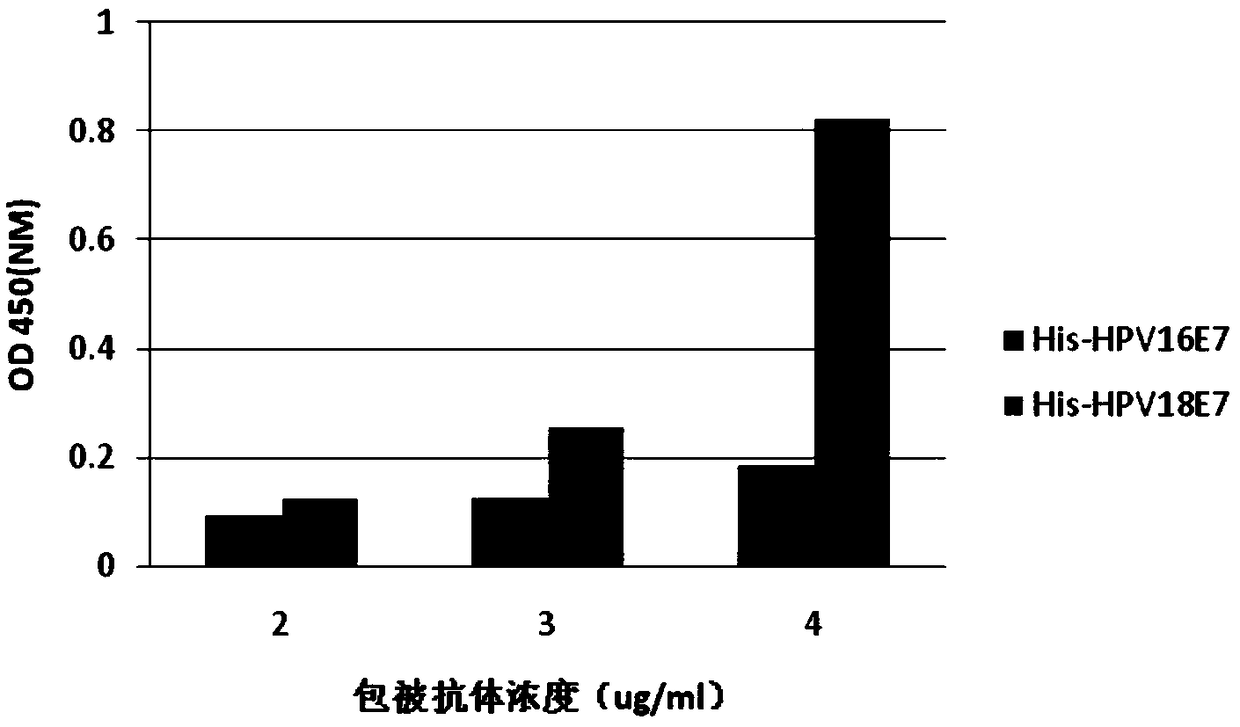
[0163] Example 2 Selection of the Concentration of Coated Antibody in Multi-antibody Coated Sandwich ELISA Detection

[0164] The mixed H11 and F1 antibodies were diluted to 2.0 μg / ml (1.0 μg / ml for H11 and F1), 3.0 μg / ml (1.5 μg / ml for H11 and F1) and 4.0 μg / ml (1.5 μg / ml for H11 and F1, respectively). F1 concentrations were 2.0 μg / ml) for coating, recombinant His-HPV16E7, His-HPV18E7 protein concentrations were 2 μg / ml as antigens, the concentrations of detection antibodies H11-Bio and F1-Bio were 1 μg / ml, each sample Set up two duplicate wells, 100 μl / well, and detect according to the steps of Example 1, such as image 3 As shown, when the total coating concentration of H11 and F1 is 4 μg / ml, that is, when the respective concentrations of the two are 2 μg / ml, negative and positive proteins can be clearly distinguished. Therefore, the total concentration of H11 and F1 antibody coating was selected to be 4 μg / ml for subsequent experiments.


PUM

PropertyMeasurementUnit
concentrationaaaaaaaaaa

Abstract

The present invention provides a method and device for detecting an immunogen. Specifically, the method provided by the present invention includes the steps of: contacting an immobilized first detection antibody with the immunogen in a sample to form a first complex; The first complex is contacted with the second detection antibody to form a second complex; the second complex is detected; the first detection antibody contains at least 2 kinds of monoclonal antibodies against the immunogen; the second detection Antibodies are labeled for detection. The present invention also provides a detection plate and a kit according to the above method. The method of the present invention can reduce the requirement for the affinity of the monoclonal antibody in the traditional ELISA method, so that the monoclonal antibody with specificity and low affinity that cannot be used in the conventional ELISA method can also be used for the detection of the antigen.

Description

technical field [0001] The invention belongs to the field of biomedicine, in particular, the invention relates to a method and device for detecting immunogen. Background technique [0002] Cervical cancer is the second most common malignant tumor in women in the world. According to statistics, there were about 530,000 new cases in the world in 2008, 85% of which occurred in developing countries. Clinical trials, molecular biology studies and etiological studies have shown that persistent infection of high-risk HPVs is a major factor in cervical cancer [zur Hausen H. Papillomaviruses and cancer: from basic studies to clinical application. Nat Rev Cancer 2002; 2:342– 50], while 16 / 18 is the most common type of high-risk HPV, and more than 2 / 3 of cervical cancers are related to its infection. [0003] After the persistent infection of HPV virus, the viral DNA integrates into the human genome and expresses oncogenic proteins (HPV oncoproteins, HPV proteins) in the host cells. ...

Claims


Application Information

Patent Timeline
Patent Type & Authority Patents(China)
IPC IPC(8): G01N33/68
Inventor 常小迦施丽君
Owner ATTOGEN BIOMEDICAL SUZHOU INC
Why Eureka
Try Eureka