Methods of reversibly binding a biotin compound to a support

a biotin compound and support technology, applied in the field of reversible binding a biotin compound to a support, can solve the problems of limited use, general undesirable, slow and complex approach, and less suitable for avidin than streptavidin

Inactive Publication Date: 2008-10-16
LIFE TECH AS
View PDF20 Cites 68 Cited by
  • Summary
  • Abstract
  • Description
  • Claims
  • Application Information

AI Technical Summary

Benefits of technology

[0036]Aspects of the present invention provide a simple method of reversal of the binding between a biotinylated moiety and a biotin-binding compound like streptavidin (or avidin) without destroying the native conformation of the target or the native status of the isolated cells. The strong interaction between streptavidin or avidin-biotin can be made much weaker by using a combination of nitro-streptavidin or avidin and modified biotin like desthiobiotin or a derivative thereof like DSB-X Biotin. A protein, such as an antibody directed against a cell-surface antigen may be biotinylated with the modified biotin. This antibody is captured or bound to the nitro-streptavidin or avidin which again is immobilized on a solid surface. This solid-phase / antibody complex is used to capture specific cells from a complex mixture. The cells with the bound antibody is then released under very gentle and fast conditions by contacting the complex with an effective amount of free biotin. The liberated cells with the biotinylated antibodies can thereby be isolated and purified.
[0037]A novel aspect of the present method over the prior art is that both parts of the biotin-streptavidin binding pair is modified to have substantially reduced affinity for each other and, as a result, the speed of release becomes much faster than what is reported in the prior art. The method avoids the harsh chemical denaturing conditions that have been previously used. It also avoids any lengthy incubation with displacement ligand, or the dilution effect occurring when eluting isolated target from affinity columns. Isolated cells will be closer to their native state than if isolated using previously available methods.

Problems solved by technology

Although the formation of such linkages between entities allows for such isolation, purification or immobilization, problems often arise if it is desired to further manipulate the isolated or purified cell population by breaking or reversing the linkage.
These non-specific interactions make avidin less suitable than streptavidin for many applications.
Such conditions are generally harmful to any bound moiety, but they work for nucleic acids as the upper temperature where the nucleic acids are damaged is about 300° C. The use of such conditions to reverse the biotin-streptavidin linkage is therefore generally undesirable, especially in the purification of proteins or separation of cells, bacteria and viruses etc. when it is important to preserving the cells integrity and maintain viability or infectivity.
This complex approach is slow and of limited use since thiols normally disrupt native protein disulfide bonds.
The drawback to this system is that binding requires a pH of 9.5 or above, while the complete dissociation of NHS-iminobiotin from streptavidin requires a pH of less than 4.
Thus, the use of NHS-iminobiotin is not suited for isolation of native cells.
Despite advances made to date, there still exists a need for new and improved methods for selectively isolating and releasing cells and other biomolecules, None of the previously reported methods for the reversible binding between biotin and streptavidin are optimal for use for the isolation of conformation sensitive targets or native cells.

Method used


Image

  • Methods of reversibly binding a biotin compound to a support
  • Methods of reversibly binding a biotin compound to a support
  • Methods of reversibly binding a biotin compound to a support

Examples


example 1

BiaCore Data of Desthiobiotin-X / Streptavidin (DSB-X / SA) Interactions

[0181]Materials and Methods

[0182]1. Immobilization Procedure:

[0183]CM5 chips (BiaCore®, BiaCore AB, Uppsala, Sweden) were activated for binding of ligands such as streptavidin or CaptAvidin by using an Amine Coupling Kit from BiaCore. The procedure was followed as provided by the manufacturer. Shortly, EDC / NHS were exposed to the CM5 chip with a flow of 5 μl / minute for 7 minutes. The ligands were then exposed to the activated CM5 surface with a flow of 5 μl / minute for 7 minutes. The excessive reactive groups were deactivated by ethanolamine with a flow of 5 μl / minute for 7 minutes. The RU of ligands were in the order between 4000-19000 RU.

[0184]2. Binding of Analytes:

[0185]The CM5 chip with covalently bound ligand (either recombinant core streptavidin or CaptAvidin (Nitroavidin)) were exposed to analytes (either biotinylated or DSB-X™ biotinylated antibodies) with a flow of 5 μl / minute at a concentration of 50-400 μ...

example 2

Procedure for Nitration of Streptavidin and Coupling to Dynabeads

[0192]Materials and Methods

[0193]1. Preparation of Nitro-Streptavidin

[0194]Streptavidin (50 mg in 5 ml of 50 mM Tris buffer, pH 8.5) was treated with 60 mM tetranitromethane for 2 hours at 25° C. Nitro-streptavidin was purified by a NAP™-25 column (GE Healthcare Life Sciences).

[0195]2. Preparation of Nitro-Streptavidin Beads

[0196]100 mg Dynabeads® M-280 Tosylactivated was washed with 0.1 M phosphate buffer, pH 7.4 (3×3 ml) and resuspended in 1.8 ml 0.1 M phosphate buffer, pH 7.4. 200 μl nitro-streptavidin (10 mg / ml in PBS) followed by 1 ml 3 M (NH4)2SO4 in 0.1 M phosphate buffer, pH 7.4 were added to the beads. After 16 hours at 37° C. on a roller, the beads were washed with 3 ml 50 mM citrate / phosphate-buffer pH 4.0 and resuspended in 3 ml. 100 μl biotin (10 mg / ml in DMSO) were added to block the unmodified biotin binding sites. After 30 minutes, the beads were washed with 3 ml 50 mM carbonate buffer pH 10 to release ...

example 3

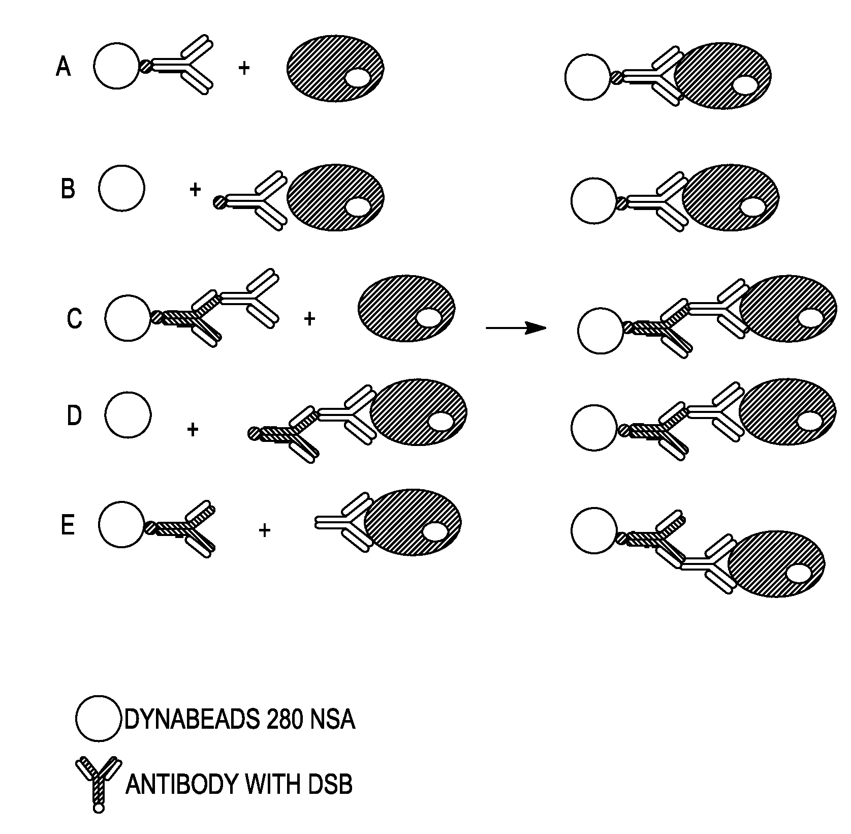
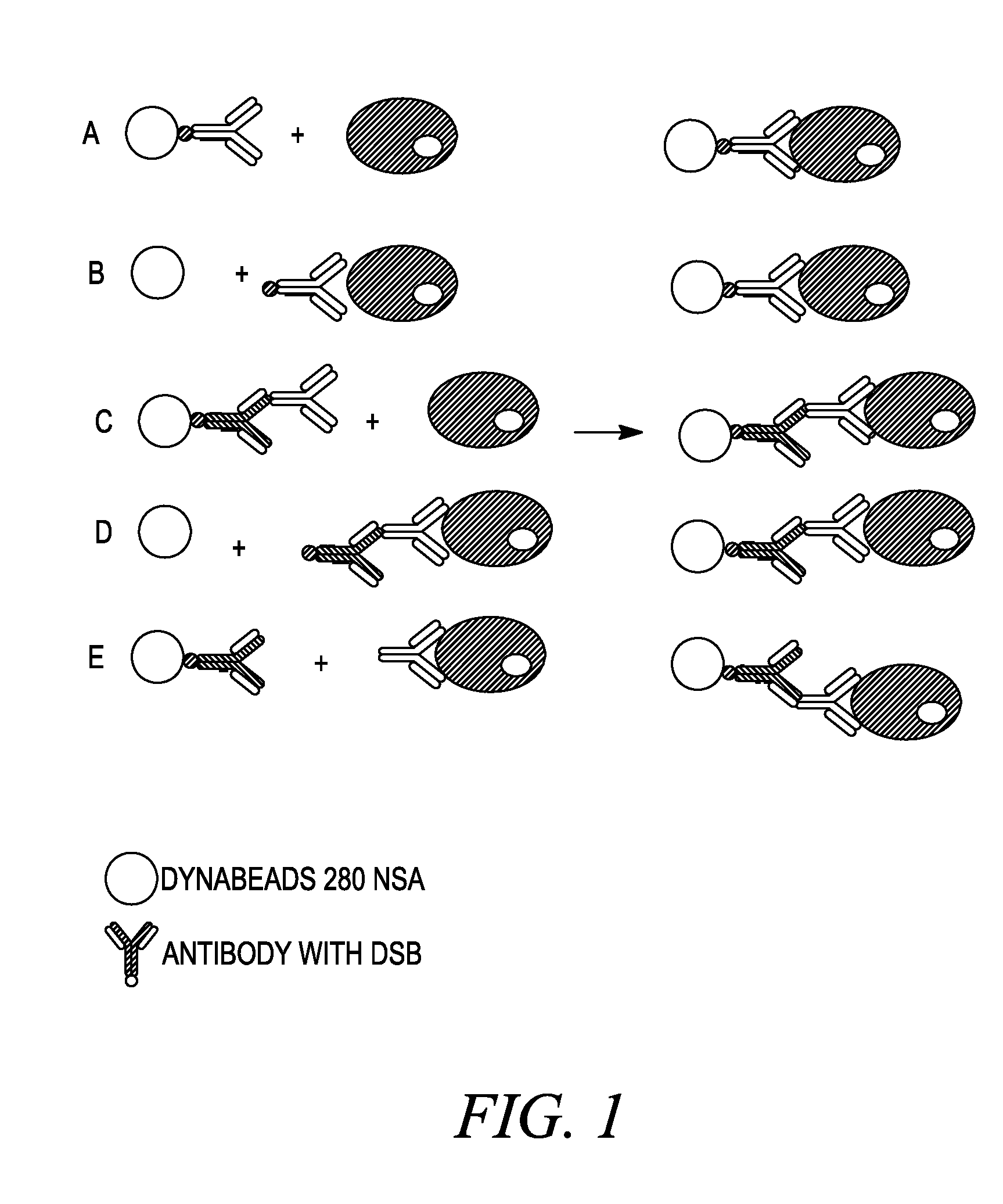
Methods Used in Different Cell Isolation Procedures as Shown in FIG. 1

[0225]Materials and Methods

[0226]Antibodies used were mouse anti-CD45 antibody, clone EO1, either DSB-labeled or not, and human anti-mouse IgG antibodies, clone HAM6, DSB-labeled.

[0227]I) Culture and Washing of Cells:

[0228]1. Grow Daudi cells in RPMI 1640, 10% FCS and 1% Na-pyruvate at 0.3-0.5×10⁶ cells / ml. Split 24 hours before use.

[0229]2. Wash cells in was-buffer (DPBS, 0.1% BSA and 2 mM EDTA). Centrifuge at 300×g for 8 minutes at 2-8° C. Resuspend in wash-buffer at 107-108 cells / ml.

[0230]II) Sensitize Cells with Antibody:

[0231]1. Add 0.5 μg antibody (anti-CD45 antibody, either DSB-labeled or not) per 106 cells to washed cells at 107-108 cells / ml

[0232]2. Incubate at 2-8° C. for 10-15 minutes (or on ice for 30-45 minutes)

[0233]Wash cells once by adding 10× excess volume of wash-buffer, centrifuge at 300×g for 8 minutes at 2-8° C. Resuspend cells in wash-buffer at 107-108 cells / ml.

[0234]3. Alternative: cells are ...


PUM

PropertyMeasurementUnit
pHaaaaaaaaaa
temperatureaaaaaaaaaa

Abstract

Methods of reversal of the binding between a biotin compound and a biotin-binding compound are disclosed. A method of reversibly releasing a biotinylated moiety from a streptavidin (or avidin) coated support is shown as an example. The strong interaction between streptavidin or avidin-biotin is made much weaker by using a combination of modified streptavidin or avidin and modified biotin like desthiobiotin or a derivative thereof like DSB-X Biotin. A protein, such as an antibody may be biotinylated with the modified biotin. When this protein is isolated by binding the modified biotin to the modified streptavidin or avidin bound to an solid surface, it may be released under very gently and very rapid conditions by addition of free biotin. In contrast to proteins obtained by the prior art release methods the protein obtained using the previously available release methods, the proteins obtained using the methods disclosed herein will maintain their native conformation. Uses of the methods in various procedures including cell detachment procedures and techniques of detection, identification, determination, purification, separation and / or isolation of target proteins or nucleic acid molecules are also described.

Description

CROSS REFERENCE TO RELATED APPLICATIONS[0001]The present application claims priority to U.S. Provisional Patent Application Ser. No. 60 / 866,021, filed Nov. 15, 2006, the contents of which are incorporated herein by reference.FIELD OF THE INVENTION[0002]The present invention provides a gentle method of reversal of the binding between a biotin compound and a biotin-binding compound. In particular, the invention relates to a method of reversibly releasing a modified biotinylated moiety from a nitro-streptavidin (or avidin) coated support.DESCRIPTION OF RELATED ART[0003]There is a continuous need in medical practice, research and diagnostic procedures for rapid, accurate, isolation or quantitative determination of different types of cells from various biological fluids.[0004]A commonly used method to achieve such isolation or determination is through the formation of a linkage between two entities, an isolation or detection entity and the target. Such linkage is often accomplished using...

Claims


Application Information

Patent Timeline
Patent Type & Authority Applications(United States)
IPC IPC(8): C40B50/18
CPCG01N33/543G01N33/82C40B50/14C12M23/20C12M47/02C12N13/00G01N33/54326
Inventor NEURAUTER, AXL A.NORDERHAUG, LARSBREKKE, OLE HENRIKSONGE, PAL
Owner LIFE TECH AS
Why Eureka
Try Eureka