Bone transport device used for massive bone defects

A handling device and segmental bone technology, which is applied in the field of devices for the treatment of large segmental bone defects, can solve the problems of poor mechanical stability of single-rod external fixation brackets, the inability to bear early weight on the affected limb, and the inability to adjust the line of force, etc., so as to avoid bad line of force , convenient clinical care, and good patient compliance

Inactive Publication Date: 2016-08-24
AFFILIATED YONGCHUAN HOSPITAL OF CHONGQING MEDICAL UNIV
View PDF9 Cites 0 Cited by
  • Summary
  • Abstract
  • Description
  • Claims
  • Application Information

AI Technical Summary

Problems solved by technology

However, the mechanical stability of the single-rod external fixator is poor, the line of force cannot be adjusted, and the affected limb cannot bear weight in the early stage. In the later stage, complications such as poor line of force, disuse osteoporosis, refracture, delayed bone union or even nonunion may occur.

Method used


Image

  • Bone transport device used for massive bone defects
  • Bone transport device used for massive bone defects
  • Bone transport device used for massive bone defects

Examples


Embodiment 1

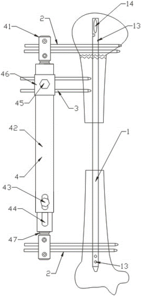
[0042] Example 1 Figure 1 to Figure 9 As shown, it is suitable for the removal of a large segment of tibia bone defect. According to different bone conditions of the patient, the intramedullary main nail can be set into three types.

[0043] The first is for patients with osteoporosis, such as Figure 1 to Figure 4 As shown, the present invention includes an intramedullary main nail 1 for axially driving bone. The intramedullary main nail 1 has an amplitude that is consistent with the anatomical structure of the human tibia, and the two ends are provided with common fixation screws. Locking hole 13. The two ends of the intramedullary main nail 1 are provided with fixing screw holes 11, and the middle part is provided with a long carrying hole 12 along the axial direction; it also includes a fixing bracket 4 and a number of fixing screws 2; the rear end of the fixing screw 2 is connected to The fixing bracket 4 is connected, and the front end is matedly connected with the fixing ...

Embodiment 2

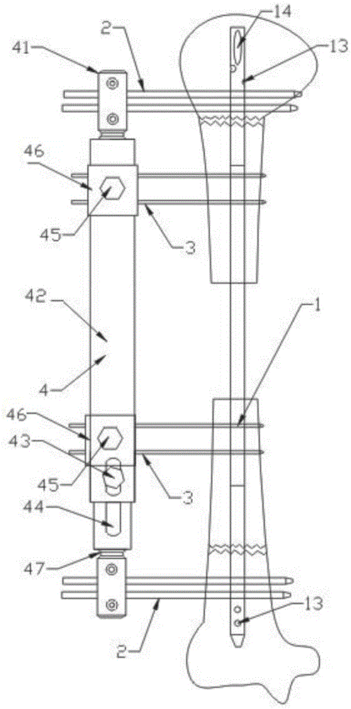
[0051] Example 2 such as Figure 10 to Figure 18 As shown, it is suitable for the removal of large segmental bone defects of the humerus. According to different bone conditions of the patient, the intramedullary main nail can be set into three types.

[0052] The first is for patients with osteoporosis, such as Figure 10 to Figure 13 As shown, the present invention includes an intramedullary main nail 1 for axially driving bone. The intramedullary main nail 1 has an amplitude that is consistent with the anatomical structure of the human humerus, and has two ends for matching with ordinary fixation screws. Locking hole 13; its proximal end is also provided with a knife hole 14 and a spiral blade 15 matching the knife 14 hole. The two ends of the intramedullary main nail 1 are provided with fixing screw holes 11, and the middle part is provided with a long carrying hole 12 along the axial direction; it also includes a fixing bracket 4 and a number of fixing screws 2; the rear end o...

Embodiment 3

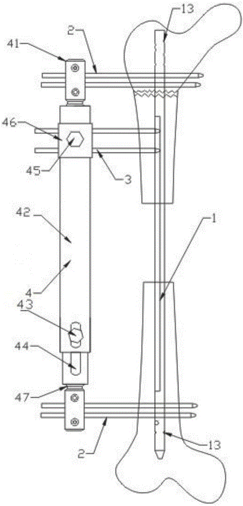
[0060] Example 3 such as Figure 19 to Figure 27 As shown, it is suitable for the removal of femoral bone defects. According to the different bone conditions of the patient, the intramedullary main nail can be set into three types.

[0061] The first is for patients with osteoporosis, such as Figure 19 to Figure 22 As shown, the present invention includes an intramedullary main nail 1 for axially driving bone. The two ends of the intramedullary main nail 1 are provided with fixing screw holes 11, and the middle of the intramedullary main nail 1 is provided with a long carrying hole 12 along the axial direction. The main intramedullary nail 1 is straight, with locking holes 13 for cooperating with ordinary fixing screws at both ends; all the locking holes 13 and fixing screw holes 11 are located on the same plane; the main intramedullary nail 1 is also An oblique locking hole 13 is provided, and the included angle with the main intramedullary nail 1 is 60° or 120°. It also includ...


PUM


Abstract

The invention discloses a bone transport device used for massive bone defects. The device comprises an intramedullary main nail used for being beaten into sclerotin in the axial direction, and the two ends of the intramedullary main nail are provided with fixing screw holes. The device further comprises a fixing support and a plurality of fixing screws. The rear ends of the fixing screws are connected with the fixing support, and the front ends of the fixing screws are matched and connected with the fixing screw holes in the intramedullary main nail. The device further comprises a plurality of transport needles capable of moving in the axial direction of the intramedullary main nail, one end of each transport needle is implanted into a bone block, and the other end of each transport needle is connected to the fixing support.

Description

Technical field [0001] The invention relates to a device for treating a large segment of bone defect by using a bone transport technique. Background technique [0002] A large segment of long bone defect refers to a bone defect in which the fracture cannot heal by itself or can only regenerate 10%, generally 2-3 times the diameter of the long bone involved. Large-scale bone defects are usually caused by high-energy trauma, infection, tumors, etc., and are often accompanied by limb shortening, deformity, osteomyelitis, muscle atrophy and adjacent joint stiffness. Repair and treatment has always been one of the biggest problems in orthopedics. According to statistics, in Germany, bone transplantation is one of the top 50 treatments most commonly received by patients; in the United States, there are about 800,000 patients who need bone transplantation each year; in China, more than the number of bone transplants required for trauma alone each year 3 million cases; large segmental b...

Claims


Application Information

Patent Timeline
Patent Type & Authority Applications(China)
IPC IPC(8): A61B17/72A61B17/66A61B17/88
CPCA61B17/66A61B17/72A61B17/8852A61B2017/564
Inventor 马坤龙栾和旭栾富钧杨帆
Owner AFFILIATED YONGCHUAN HOSPITAL OF CHONGQING MEDICAL UNIV
Why Eureka
Try Eureka