Highlight region eliminating method of endoscopic image

An image and area technology, which is applied in the field of medical endoscopic image highlight removal, can solve the problems of difficult to achieve natural restoration and natural restoration of endoscopic images, and achieve the effect of accurate detection and segmentation

Active Publication Date: 2016-09-21
BEIJING INSTITUTE OF TECHNOLOGYGY
View PDF4 Cites 38 Cited by
  • Summary
  • Abstract
  • Description
  • Claims
  • Application Information

AI Technical Summary

Problems solved by technology

Although the currently proposed algorithm can solve the defects of existing endoscopic image highlight pixel detection and repair algorithms to a certain extent, the effect of reflection highlight pixel extraction needs to be improved. Difficult to achieve natural restoration effect
[0005] Therefore, this field needs an effective method for eliminating the highlight area of ​​the endoscopic image to solve the problem that it is difficult to realize the natural repair of the endoscopic image when the highlight area is large

Method used


Image

  • Highlight region eliminating method of endoscopic image
  • Highlight region eliminating method of endoscopic image
  • Highlight region eliminating method of endoscopic image

Examples


Embodiment Construction

[0048] The illustrated embodiments are disclosed with reference to the drawings. It should be understood, however, that the disclosed embodiments are merely embodiments that may be shown in various and alternative forms. The figures are not necessarily to scale and some features may be exaggerated or minimized to show details of particular components. Specific structural and functional details disclosed are not to be interpreted as limiting, but as a representative basis for teaching one skilled in the art how to practice the present disclosure.

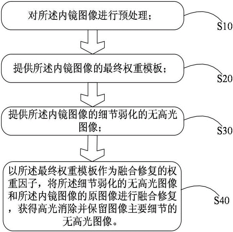
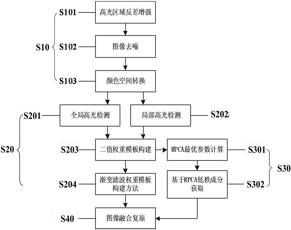
[0049]In order to overcome the deficiencies in existing endoscopic image highlight detection and repair algorithms, the present invention provides a method for eliminating highlight areas of endoscopic images based on superpixels and Otsu (OTSU) thresholds. This method is based on low-rank feature fusion. The method of eliminating the highlight area of ​​the mirror image can realize the accurate detection and segmentation of the loc...


PUM


Abstract

The present invention provides a highlight region eliminating method of an endoscopic image. The method comprises the steps of (S10) preprocessing the endoscopic image, (S20) providing the final weighting template needed by an image fusion operation, (S30) providing a no-highlight image with weakened details of the endoscopic image, (S40) with the final weight template as the weight factor of fusion repair, carrying out fusion repair of the no-highlight image with the weakened details and the original image of the endoscopic image, and obtaining the no-highlight image with the elimination of highlight and the maintenance of image main details. The invention aims to provide the highlight region eliminating method of the endoscopic image, and the natural repair of the endoscopic image can be realized when the highlight region is large.

Description

technical field [0001] The invention relates to a method for eliminating high light in medical endoscopic images, which can be used to assist doctors in better observation and disease diagnosis in the medical endoscopic imaging diagnosis and treatment process. Background technique [0002] During the endoscopic imaging process, the high-light reflections on the surface of human organs usually appear as bright areas with saturated brightness. These bright areas will reduce the image quality and have a great impact on the observation of the surgical process and the judgment of lesions, and even It becomes a source of error in many vision-based analysis algorithms and negatively affects many gradient-based computer vision algorithms. [0003] Over the past decades, a large number of algorithms have been proposed to detect and repair specular reflections in endoscopic images. Arnold, et al. based on the method of global nonlinear filtering and color threshold to detect the high...

Claims


Application Information

Patent Timeline
Patent Type & Authority Applications(China)
IPC IPC(8): G06T5/00G06T5/40
CPCG06T5/002G06T5/007G06T5/40
Inventor 杨健郜业飞王涌天艾丹妮丛伟建范敬凡
Owner BEIJING INSTITUTE OF TECHNOLOGYGY
Why Eureka
Try Eureka