Morphological segmentation-based eye fundus image lesion detection method

A technology based on morphology and fundus images, applied in the field of image processing, can solve the problems of unsatisfactory detection of hemorrhage area, speed up detection speed, and large amount of calculation, and achieve the effect of improving real-time operation, speeding up detection speed, and avoiding interference.

Inactive Publication Date: 2016-11-23
SHANGHAI JIAO TONG UNIV
View PDF0 Cites 11 Cited by
  • Summary
  • Abstract
  • Description
  • Claims
  • Application Information

AI Technical Summary

Problems solved by technology

[0016] In view of the inaccurate response to some weak edges in the prior art, the detection of hemorrhage areas connected to blood vessels is not ideal, and the calculation amount is large, and the calculation speed is insufficient, the present invention proposes a fundus based on morphological segmentation The image lesion detection method uses the gradient information, gray level information of the image and the characteristics of similar gray levels of blood vessels

Method used


Image

  • Morphological segmentation-based eye fundus image lesion detection method
  • Morphological segmentation-based eye fundus image lesion detection method
  • Morphological segmentation-based eye fundus image lesion detection method

Examples


Embodiment 1

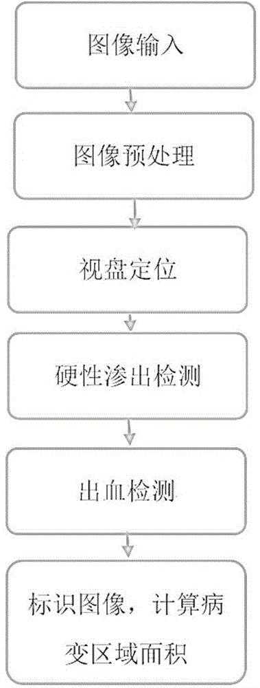
[0033] Such as figure 1 As shown, this embodiment includes the following steps:

[0034] The first step is to input the fundus image to be detected and perform preprocessing, specifically:

[0035] 1.1) Use the brightness component of HSV space to achieve brightness balance: first convert the fundus image into HSV space, and perform the following operations on the V component: where: X V Represents the V component of the pixel, X’ V Indicates the V component value of the updated pixel point; then X' V Return the RGB space of the pixel, that is, the brightness equalization is completed.

[0036] 1.2) Contrast enhancement is achieved by Constrained Contrast Adaptive Histogram Equalization (CLAHE);

[0037] This operation not only satisfies the effect of adaptive histogram equalization to redistribute the brightness through the local histogram to achieve the effect of contrast enhancement, but also overcomes the problem of excessive amplification of noise caused by ordinary...


PUM


Abstract

A fundus image lesion detection method based on morphological segmentation. First, the fundus image is smoothed and filtered, and the region growing method is used to locate the region where the optic disc is located; then the fundus background image with the exudate area removed is obtained through morphological processing, and is segmented by thresholding A mixed area image including blood vessels and hemorrhage is obtained; finally, the edge detection is performed by Kirsch operator and the blood vessel image is obtained by region growing. The invention avoids the possible interference caused by blood vessels with similar gray levels in other bleeding detection algorithms, and also speeds up the detection speed.

Description

technical field [0001] The present invention relates to a technique in the field of image processing, in particular to a detection method for hard exudation based on background restoration of a healthy region of a fundus image and a method for detection of hemorrhage based on edge detection and region growth. Background technique [0002] With the development of computer graphics processing technology, the analysis of fundus images no longer relies solely on ophthalmologists' naked eyes. out and bleed techniques, and a great deal of research has been done. Traditional manual qualitative analysis lacks quantitative means. Relying on the computer to quickly and reliably automatically identify the lesions in the fundus image can prevent doctors from injecting special drugs into patients to make the fundus image clear, and at the same time avoid the doctor's manual judgment of film reading, saving a lot of manpower, material resources and time. The implementation of lesion det...

Claims


Application Information

Patent Timeline
IPC IPC(8): G06T7/00G06F19/00
Inventor 许哲宇胡昊坤马力天殷本俊盛斌
Owner SHANGHAI JIAO TONG UNIV
Why Eureka
Try Eureka