Eureka AIR delivers breakthrough ideas for toughest innovation challenges, trusted by R&D personnel around the world.

A plug-in electronic visual lens group endoscope structure

A lens group and plug-in technology, applied in endoscopy, medical science, surgery, etc., can solve problems such as difficult to meet the high-quality image acquisition requirements of image diagnosis and image quality limitations of lens groups, so as to improve the flexibility of position adjustment , Solve the poor imaging quality and improve the effect of image acquisition quality

Active Publication Date: 2018-10-12
ZHEJIANG UE MEDICAL
View PDF6 Cites 0 Cited by
  • Summary
  • Abstract
  • Description
  • Claims
  • Application Information

AI Technical Summary

Problems solved by technology

However, in this technical solution, the ring light is surrounded by optical fibers and bundled at the root, and the camera module installed at the front of the mirror makes the setting of the lens group simple and the imaging quality is limited, making it difficult to meet the high-quality image acquisition requirements of image diagnosis.

Method used


Image

  • A plug-in electronic visual lens group endoscope structure
  • A plug-in electronic visual lens group endoscope structure
  • A plug-in electronic visual lens group endoscope structure

Examples


Embodiment Construction

[0018] Below by specific embodiment, in conjunction with accompanying drawing, the technical solution of the present invention is described in further detail:

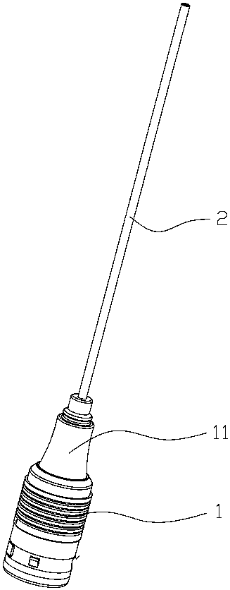
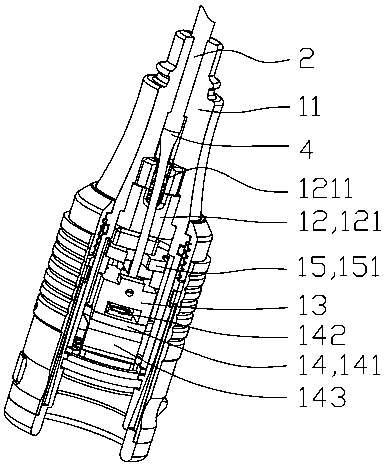
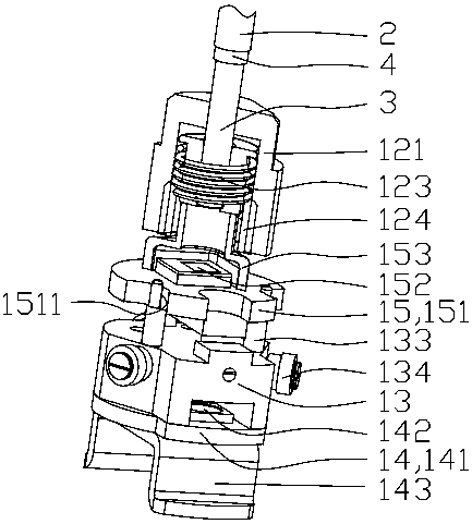
[0019] see figure 1 and figure 2 , a plug-in electronic visual lens group endoscope structure, including a plug-in part 1 plugged with the main body of the electronic visual endoscope (not shown in the figure), and a mirror housing 11 in the plug-in part 1 The mirror tube 2 connected to the socket 1, the lens group 3 arranged in the mirror tube 2, the light guide fiber 4 arranged around the lens group 3 and extending axially, and the light source module 14 arranged in the socket 1 and camera module 15. see image 3 and Figure 4 , wherein, the lens group 3 is fixed and centered on the camera chip 152 of the camera module 15 through a lens group fixing structure 12; On a fixed frame 13 , the LED light source 142 on the light source module 14 is aligned to irradiate the root of the light-guiding optical fiber 4 . ...


PUM


Abstract

The invention discloses an insertion and connection type electronic visual lens group endoscope structure, which comprises an insertion and connection part, an endoscope pipe, an image pickup module and a light source module, wherein the endoscope pipe is connected onto the insertion and connection part, and comprises lens groups arranged in the center of the endoscope pipe and a light guide optical fiber which surrounds the endoscope pipe and extends in the longitudinal direction of the endoscope pipe; the light guide optical fiber is clustered in one side at the root part of the endoscope pipe, is in bias extension and is fixed in the insertion and connection part; a light source in the light source module is correspondingly irradiated at the root part of the light guide optical fiber. Through the arrangement of a plurality of lens groups in the endoscope pipe, a high-quality optical image can be obtained, so that the image is directly received by the image pickup module arranged at the root part of the lens groups, and the high-quality optical image is obtained; meanwhile, one bias structure is used, so that the light source is annularly conveyed to the periphery of the lens from the root part of the endoscope pipe by the light guide optical fiber, so that the imaging quality of the endoscope is further improved.

Description

technical field [0001] The invention belongs to the technical field of medical devices, and in particular relates to a visualized medical device that penetrates into a human body to observe, diagnose and treat diseased organs inside the human body. Background technique [0002] With the advancement of medical device technology, endoscopes have become an essential medical device in hospitals. The introduction of modern electronic imaging technology has made the image acquisition of traditional endoscopes more convenient and improved the efficiency and quality of medical diagnosis. The Chinese invention patent application with the notification number CN105520708A published by the applicant on April 27, 2016 discloses a quick-connect electronic visual endoscope device, including a device main body, a human-computer interaction part, a control module, a power supply and the front end of the mirror inserted through the plug-in structure; the front end of the mirror is provided w...

Claims


Application Information

Patent Timeline
Patent Type & Authority Patents(China)
IPC IPC(8): A61B1/05A61B1/07A61B1/00
CPCA61B1/00163A61B1/05A61B1/0607A61B1/07
Inventor 陆静江春才陈盛袁洪文朱威灵李卫平
Owner ZHEJIANG UE MEDICAL
Why Eureka
Eureka Blog
Learn More