
Method and system for treating medical images

A medical image and image size technology, applied in the field of computer vision, can solve the problems of false positives, insufficient characterization and distinction between lesions and normal areas, and achieve the effect of improving accuracy, overcoming insufficient feature extraction, and reducing false negatives.

Inactive Publication Date: 2017-04-19
北京羽医甘蓝信息技术有限公司
View PDF0 Cites 13 Cited by
  • Summary
  • Abstract
  • Description
  • Claims
  • Application Information

AI Technical Summary

Problems solved by technology

[0004] However, the application of existing technologies in gastrointestinal endoscopy will produce a large number of false positives, mainly because the models used by such methods usually only contain a hidden layer for extracting features, and the extracted features are often insufficient. To characterize and distinguish diseased spots from normal areas

Method used


Image

  • Method and system for treating medical images
  • Method and system for treating medical images
  • Method and system for treating medical images

Examples


Embodiment Construction

[0019] Exemplary implementations of the present invention are described below in conjunction with the accompanying drawings, which include various details of the implementations of the present invention to facilitate understanding, and they should be regarded as exemplary only. Accordingly, those of ordinary skill in the art will recognize that various changes and modifications of the embodiments described herein can be made without departing from the scope and spirit of the invention. Also, descriptions of well-known functions and constructions are omitted in the following description for clarity and conciseness.

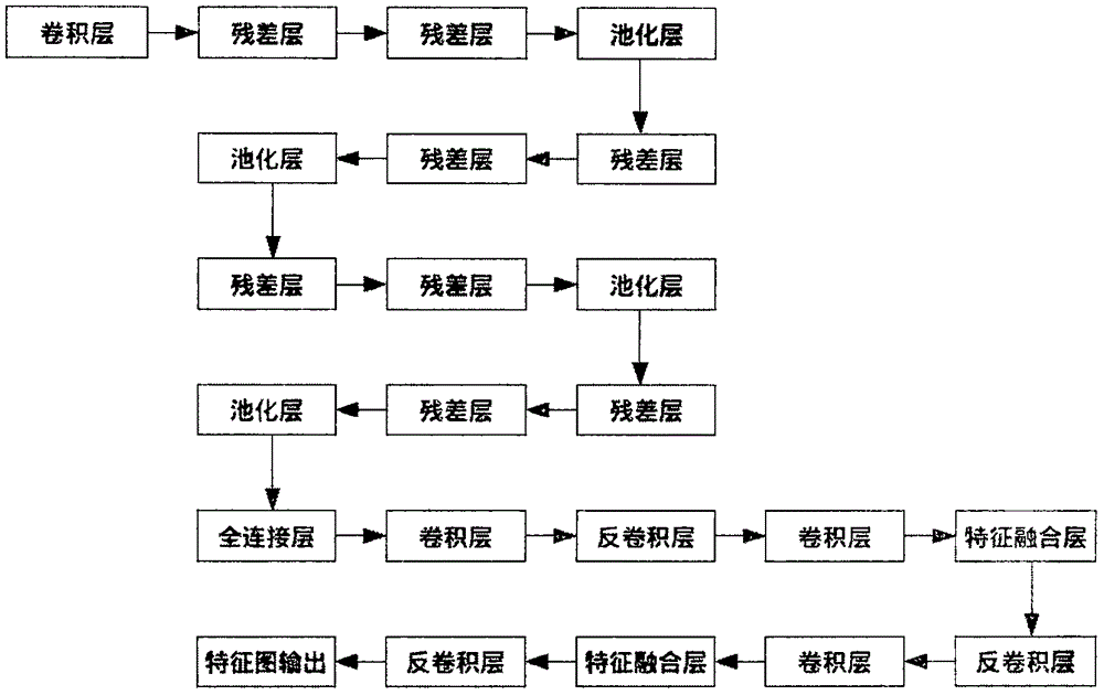
[0020] The disadvantages of the prior art have already been explained in the background art. The neural network used in the deep learning solution adopted in the technical solution of the present invention has the characteristics of extracting high-level features of objects. Since the high-level feature information is a linear and nonlinear transformation of the u...


PUM


Abstract

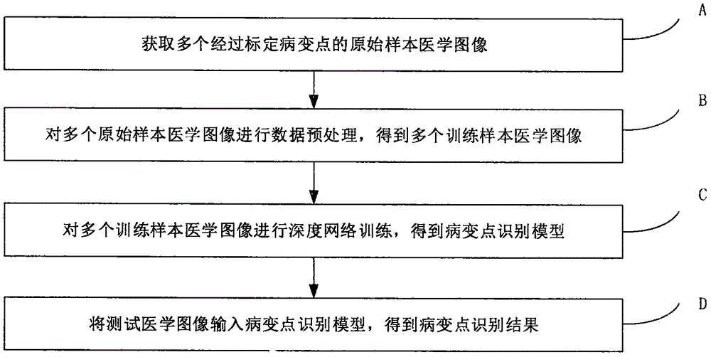
The invention provides an accurate and reliable method and an accurate and reliable system for treating medical images and aims to solve a problem in the prior art. The method comprises steps that A, multiple original sample medical images after lesion point calibration are acquired; B, data pre-treatment on the multiple original sample medical images is carried out to acquire multiple training sample medical images; C, depth neural training for the multiple training sample medical images is carried out to acquire a lesion point identification model; and D, test medical images are inputted to the lesion point identification model to acquire a lesion point identification result.

Description

technical field [0001] The invention relates to the technical field of computer vision, in particular to a method and system for processing medical images. Background technique [0002] Using algorithms to automatically identify lesions or potential lesions (such as intestinal polyps) from medical images by computers is a problem that people have been trying to solve for many years. [0003] The traditional computer automatic recognition algorithm works like this: convert the original image input (pixel value) into human-engineered features, such as SIFT, HOG features, etc. Then put these transformed features into a pre-trained shallow detector for detection. The detection process can be roughly understood as sliding a detection window with a preset size on the original image. If calculated at a certain position If the detection score is higher than a preset threshold, it is considered that there is a lesion or potential lesion of our interest in this position. [0004] Ho...

Claims


Application Information

Patent Timeline
IPC IPC(8): G06K9/62G06T5/40
CPCG06T5/40G06F18/214
Inventor 丁鹏
Owner 北京羽医甘蓝信息技术有限公司
Learn More