
Medical image segmentation method

A medical image and image technology, applied in the field of image processing, can solve problems such as single-objective optimization function, without considering the clustering effect, etc., to achieve the effect of enhanced anti-noise ability and high accuracy

Active Publication Date: 2017-04-19
JIANGSU UNIV
View PDF3 Cites 14 Cited by
  • Summary
  • Abstract
  • Description
  • Claims
  • Application Information

AI Technical Summary

Problems solved by technology

[0008] The purpose of the present invention is to propose a medical image segmentation method that fully considers the image as a whole and each component, and enables them to achieve optimal results in view of the limitations of the above-mentioned prior art, thereby effectively solving most of the clustering problems. The algorithm only has a single objective optimization function and does not consider the defects of the clustering effect of the components, which significantly improves the anti-noise ability, makes the clustered image closer to the objective and real situation of each object, and provides more accurate medical diagnosis. image based on

Method used


Image

  • Medical image segmentation method
  • Medical image segmentation method
  • Medical image segmentation method

Examples


Embodiment Construction

[0034] The medical image clustering method will be further described below mainly in conjunction with the accompanying drawings and specific embodiments.

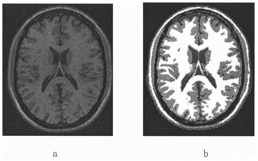
[0035] In this embodiment, the brain MRI map is selected for analysis, and the original brain MRI containing noise points is selected. figure 2 a to illustrate the corresponding results after the implementation of the present invention, the concrete steps are as follows:

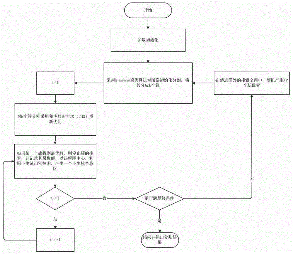
[0036] A. The computer reads the original image of the brain MRI map, sets the search scale N=100, the number of clusters K=3, the number of optimization iterations T=100 in each cluster, and the harmony search algorithm parameter memory value matrix HMCR=I , the fine-tuning probability PAR=0.01, the pitch fine-tuning bandwidth bw=0.0001, and the maximum number of iterations T max=200.

[0037]B. Using the K-means algorithm to segment the medical image and divide it into 3 clusters.

[0038] C. According to the segmentation results in B, each segmented...


PUM


Abstract

The present invention relates to a medical image segmentation method and belongs to the image processing field. The method of the present invention is based on a harmony search algorithm and is realized by the steps of pre-processing an image, extracting the characteristics of the image, pre-segmenting the image, carrying out the global optimal harmony search, carrying out the optimal harmony search in partial areas of the image, etc. The theory and practice prove that the method of the present invention properly solves the problems of a single objective function and a single threshold value and the problem of not considering the clustering effect of the components, in a clustering algorithm. After adopting the method of the present invention, not only the medical images can be segmented efficiently, but also the important image characteristics do not lose, so that the high-quality medical segmentation images can be obtained, and the diagnosis reading demands of the medical care personnel are satisfied.

Description

technical field [0001] The invention relates to an image segmentation method, in particular to a medical image segmentation method, which belongs to the field of image processing. Background technique [0002] With the rapid development of human medical imaging technologies such as Computed Tomography (CT) and MRI (Megnectic Resonance lmaging, MRI), medical images are playing an increasingly important role in clinical medical diagnosis. [0003] As an unsupervised learning algorithm, clustering is used in many practical problems due to its simplicity, ease of operation, and good robustness, such as data mining, image processing, bioinformatics, decision-making and planning, and so on. Clustering method is also one of the methods often used in image segmentation. The main idea of ​​this type of method is to divide the features into multiple categories according to the features in the image data and according to a certain clustering algorithm idea. The image features in the s...

Claims


Application Information

Patent Timeline
Patent Type & Authority Applications(China)
IPC IPC(8): G06T7/11G06K9/62
CPCG06T2207/10088G06T2207/30016G06F18/23213
Inventor 刘哲宋余庆包翔
Owner JIANGSU UNIV
Learn More